PY17X_1231100 adenylyl cyclase beta, putative (ACbeta)
Disruptability [+]
| Species | Disruptability | Reference | Submitter | |
|---|---|---|---|---|
| P. berghei ANKA |
Refractory |
PlasmoGEM (Barseq) | PlasmoGEM | |
| P. falciparum 3D7 |
Refractory |
USF piggyBac screen (Insert. mut.) | USF PiggyBac Screen | |
Mutant phenotypes [+]
| Species | Stage | Phenotype | Reference | Submitter |
|---|---|---|---|---|
| P. falciparum 3D7 | Asexual |
Egress defect |
31075098 Delayed egress |
Theo Sanderson, Google AI |
| P. falciparum 3D7 | Asexual |
Invasion defect |
31075098 | Theo Sanderson, Google AI |
Imaging data (from Malaria Metabolic Pathways)
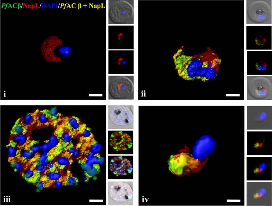
Expression of PfACb in P. falciparum blood stages. Immunofluorescence assays (IFA) were used to detect PfACb (green) in P. falciparum rings (i), trophozoites (ii), schizonts (iii) and merozoites (iv) using mouse antisera against a peptide derived from PfACb. Nuclear DNA was stained with DAPI (blue). Rabbit antiserum against P. falciparum cytoplasmic protein PfNAPL (red) was used for co-localization. Yellow indicates overlap of PfACb and PfNAPL. PfACb is thus expressed in P. falciparum merozoites and is localized in the cytoplasm.Dawn A, Singh S, More KR, Siddiqui FA, Pachikara N, Ramdani G, Langsley G, Chitnis CE. The Central Role of cAMP in Regulating Plasmodium falciparum Merozoite Invasion of Human Erythrocytes. PLoS Pathog. 2014 18;10(12):e1004520.
See original on MMP
Upper panel: Location of Dansyl-MAS 7 and MAS 17 peptides, within the parasite infected erythrocyte. Parasite nuclei are shown in blue, Dansyl fluorescence in green and BODIPY-TR-Ceramide in red. A merge and bright field (BF) image are shown. Scale bar = 3 μm. MAS-7 probably binds to adenylyl cyclase .Lower panel: Location of Dansyl-MAS 7 peptide, within the parasite infected erythrocyte following 1 hour (upper panel) and 2 hour (lower panel) incubation. Scale bar = 3 μm.Peatey CL, Dixon MW, Gardiner DL, Trenholme KR. Temporal evaluation ofcommitment to sexual development in Plasmodium falciparum. Malar J. 2013 12:134.
See original on MMP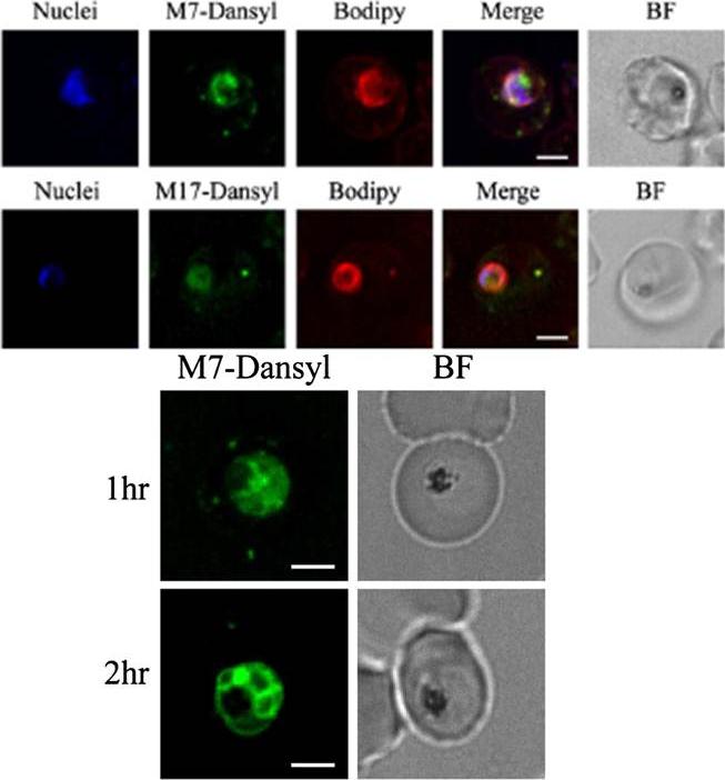
Upper panel: Location of Dansyl-MAS 7 and MAS 17 peptides, within the parasite infected erythrocyte. Parasite nuclei are shown in blue, Dansyl fluorescence in green and BODIPY-TR-Ceramide in red. A merge and bright field (BF) image are shown. Scale bar = 3 μm. MAS-7 probably binds to adenylyl cyclase .Lower panel: Location of Dansyl-MAS 7 peptide, within the parasite infected erythrocyte following 1 hour (upper panel) and 2 hour (lower panel) incubation. Scale bar = 3 μm.Peatey CL, Dixon MW, Gardiner DL, Trenholme KR. Temporal evaluation ofcommitment to sexual development in Plasmodium falciparum. Malar J. 2013 12:134.
See original on MMPMore information
| PlasmoDB | PY17X_1231100 |
| GeneDB | PY17X_1231100 |
| Malaria Metabolic Pathways | Localisation images Pathways mapped to |
| Previous ID(s) | null |
| Orthologs | PBANKA_1227600 , PCHAS_1228300 , PF3D7_0802600 , PKNH_0116300 , PVP01_0117600 , PVX_093610 |
| Google Scholar | Search for all mentions of this gene |