PF3D7_1147800 membrane associated erythrocyte binding-like protein (MAEBL)
Disruptability [+]
| Species | Disruptability | Reference | Submitter |
|---|---|---|---|
| P. falciparum 3D7 |
Possible |
28371168 Sproozoites don't invade salivary glands, those isolated from hemolymph are defective in cell-traversal and invasion |
Theo Sanderson, Wellcome Trust Sanger Institute |
| P. falciparum 3D7 |
Possible |
USF piggyBac screen (Insert. mut.) | USF PiggyBac Screen |
| P. falciparum 3D7 |
Possible |
15811533 | Theo Sanderson, Francis Crick Institute |
Mutant phenotypes [+]
| Species | Stage | Phenotype | Reference | Submitter |
|---|---|---|---|---|
| P. falciparum 3D7 | Asexual |
No difference |
28371168 Sproozoites don't invade salivary glands, those isolated from hemolymph are defective in cell-traversal and invasion |
Theo Sanderson, Wellcome Trust Sanger Institute |
| P. falciparum 3D7 | Asexual |
Possible |
15811533 | Theo Sanderson, Francis Crick Institute |
| P. falciparum 3D7 | Gametocyte |
No difference |
28371168 Sproozoites don't invade salivary glands, those isolated from hemolymph are defective in cell-traversal and invasion |
Theo Sanderson, Wellcome Trust Sanger Institute |
| P. falciparum 3D7 | Ookinete |
No difference |
28371168 Sproozoites don't invade salivary glands, those isolated from hemolymph are defective in cell-traversal and invasion |
Theo Sanderson, Wellcome Trust Sanger Institute |
| P. falciparum 3D7 | Oocyst |
No difference |
28371168 Sproozoites don't invade salivary glands, those isolated from hemolymph are defective in cell-traversal and invasion |
Theo Sanderson, Wellcome Trust Sanger Institute |
| P. falciparum 3D7 | Sporozoite |
Attenuated |
28371168 Sproozoites don't invade salivary glands, those isolated from hemolymph are defective in cell-traversal and invasion |
Theo Sanderson, Wellcome Trust Sanger Institute |
Imaging data (from Malaria Metabolic Pathways)

MAEBL localized with rhoptry proteins and on the merozoite surface and not with microneme proteins. Dual-label indirect immunofluorescence assays analyzed by scanning confocal laser microscopy are shown in each row of images, using primary antibodies against a rhoptry and microneme protein. In each row, the immunolocalization pattern for the first protein listed is in green in the first left-hand panel and the second protein listed is the second panel shown in red. The third panel is the merged image of the first two panels, the final right-hand panel is the brightfield image. Color was assigned electronically. Markers: RhopH2 PFI1445w; EBA-175 MAL7P1.176; RAP1 PFE0080c.Blair PL, Kappe SH, Maciel JE, Balu B, Adams JH. Plasmodium falciparum MAEBL is a unique member of the ebl family. Mol Biochem Parasitol. 2002 122:35-44. Copyright Elsevier 2009.
See original on MMP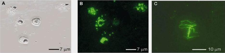
Phase contrast photograph of P. falciparum schizonts and free merozoites (100x) showing the reactivity of the mouse polyclonal anti-r-PfM2. (B) IFA reactivity of the mouse anti-r-PfM2 serum with methanol fixed late stage schizonts and free merozoites of P. falciparum 3D7 clone (100x). (C) IFA reactivity to air dried P. falciparum sporozoites (100x) using mouse anti-r-PfM2 antibody. Apical punctate fluorescence was observed in free merozoites and in late-stage schizonts. Positive fluorescence was also recorded on the surface of anopheles salivary gland sporozoites.Ghai M, Dutta S, Hall T, Freilich D, Ockenhouse CF. Identification, expression, and functional characterization of MAEBL, a sporozoite and asexual blood stage chimeric erythrocyte-binding protein of Plasmodium falciparum. Mol Biochem Parasitol. 2002 123:35-45. Copyright Elsevier
See original on MMPMore information
| PlasmoDB | PF3D7_1147800 |
| GeneDB | PF3D7_1147800 |
| Malaria Metabolic Pathways | Localisation images Pathways mapped to |
| Previous ID(s) | PF11_0486, PF3D7_1147800.1, PF3D7_1147800.2 |
| Orthologs | PBANKA_0901300 , PBANKA_0901300 , PCHAS_0702500 , PKNH_0945700 , PVP01_0948400 , PVX_092975 |
| Google Scholar | Search for all mentions of this gene |