PF3D7_1404600 adenylyl cyclase alpha (ACalpha)
Disruptability [+]
| Species | Disruptability | Reference | Submitter | |
|---|---|---|---|---|
| P. falciparum 3D7 |
Possible |
USF piggyBac screen (Insert. mut.) | USF PiggyBac Screen | |
| P. berghei ANKA |
Possible |
RMgm-235 | Imported from RMgmDB | |
Mutant phenotypes [+]
| Species | Stage | Phenotype | Reference | Submitter |
|---|---|---|---|---|
| P. berghei ANKA | Asexual |
No difference |
RMgm-235 | Imported from RMgmDB |
| P. berghei ANKA | Gametocyte |
No difference |
RMgm-235 | Imported from RMgmDB |
| P. berghei ANKA | Ookinete |
No difference |
RMgm-235 | Imported from RMgmDB |
| P. berghei ANKA | Oocyst |
No difference |
RMgm-235 | Imported from RMgmDB |
| P. berghei ANKA | Sporozoite |
Difference from wild-type |
RMgm-235
Normal numbers of salivary gland sporozoites are formed. Gliding motility of sporozoites was comparable to that of wild type parasites. The cell-traversal activity of mutant sporozoites was slightly lower, but not significantly different from wild type sporozoites.The mutant sporozoites are approximately 50% less infective to Hepa1-6 cells and to C57Bl6 mice than wild type sporozoites. |
Imported from RMgmDB |
| P. berghei ANKA | Liver |
Difference from wild-type |
RMgm-235
Gliding motility of sporozoites was comparable to that of wild type parasites. The cell-traversal activity of mutant sporozoites was slightly lower, but not significantly different from wild type sporozoites.The mutant sporozoites are approximately 50% less infective to Hepa1-6 cells and to C57Bl6 mice than wild type sporozoites. |
Imported from RMgmDB |
Imaging data (from Malaria Metabolic Pathways)

Upper panel: Location of Dansyl-MAS 7 and MAS 17 peptides, within the parasite infected erythrocyte. Parasite nuclei are shown in blue, Dansyl fluorescence in green and BODIPY-TR-Ceramide in red. A merge and bright field (BF) image are shown. Scale bar = 3 μm. MAS-7 probably binds to adenylyl cyclase .Lower panel: Location of Dansyl-MAS 7 peptide, within the parasite infected erythrocyte following 1 hour (upper panel) and 2 hour (lower panel) incubation. Scale bar = 3 μm.Peatey CL, Dixon MW, Gardiner DL, Trenholme KR. Temporal evaluation ofcommitment to sexual development in Plasmodium falciparum. Malar J. 2013 12:134.
See original on MMP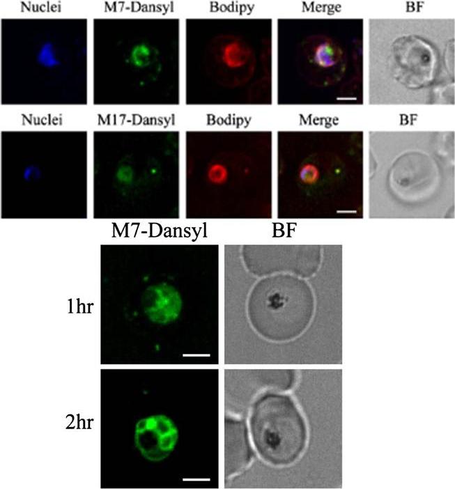
Upper panel: Location of Dansyl-MAS 7 and MAS 17 peptides, within the parasite infected erythrocyte. Parasite nuclei are shown in blue, Dansyl fluorescence in green and BODIPY-TR-Ceramide in red. A merge and bright field (BF) image are shown. Scale bar = 3 μm. MAS-7 probably binds to adenylyl cyclase .Lower panel: Location of Dansyl-MAS 7 peptide, within the parasite infected erythrocyte following 1 hour (upper panel) and 2 hour (lower panel) incubation. Scale bar = 3 μm.Peatey CL, Dixon MW, Gardiner DL, Trenholme KR. Temporal evaluation ofcommitment to sexual development in Plasmodium falciparum. Malar J. 2013 12:134.
See original on MMPMore information
| PlasmoDB | PF3D7_1404600 |
| GeneDB | PF3D7_1404600 |
| Malaria Metabolic Pathways | Localisation images Pathways mapped to |
| Previous ID(s) | PF14_0043, PF14_0788, PF3D7_1404600.1, PF3D7_1404600.2, PF3D7_1404600.3 |
| Orthologs | PBANKA_1037500 , PCHAS_1038300 , PKNH_1353500 , PVP01_1344200 , PVX_086205 , PY17X_1039900 |
| Google Scholar | Search for all mentions of this gene |