PY17X_1109000 triose phosphate transporter, putative (TPT)
Disruptability [+]
| Species | Disruptability | Reference | Submitter | |
|---|---|---|---|---|
| P. berghei ANKA |
Refractory |
23041242 | Theo Sanderson, Wellcome Trust Sanger Institute | |
| P. falciparum 3D7 |
Refractory |
USF piggyBac screen (Insert. mut.) | USF PiggyBac Screen | |
Mutant phenotypes [+]
| Species | Stage | Phenotype | Reference | Submitter |
|---|---|---|---|---|
| P. falciparum 3D7 | Asexual |
Attenuated |
32815516 (Conditional)
Conditional system in which mutant was generated with supplementation so that apicoplast is not required. "Upon removal of mevalonate both deletion lines failed to grow " |
Theo Sanderson, Francis Crick Institute |
Imaging data (from Malaria Metabolic Pathways)
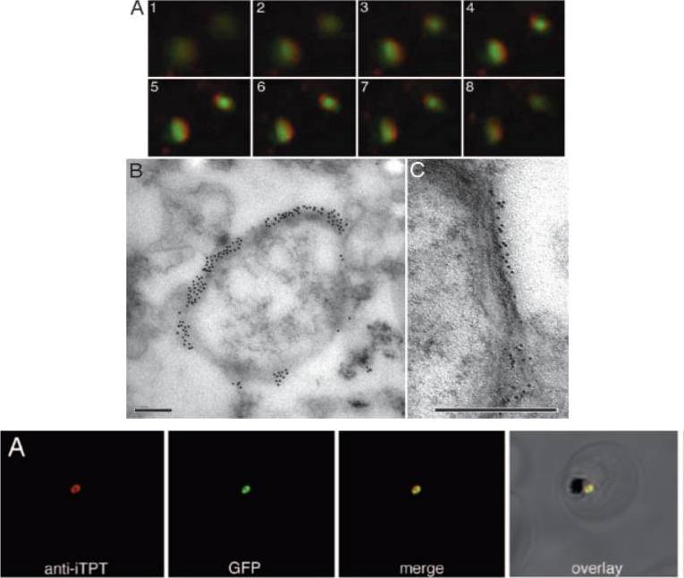
Immunolocalization of PfoTPT to the outside of free, intact apicoplasts. (A) Z-stack of immunofluorescent images (1– 8) showing localization of anti-PfoTPT (red) around apicoplast-targeted GPF (green) in parasites expressing PfACP(leader)-GFP. (B) Electron micrograph of intact apicoplast labeled with anti-PfoTPT antibodies and 20-nm colloidal gold. (C) Tannic acid fixation showing four bounding membranes around isolated apicoplast labeled with anti-PfoTPT antibodies and 10-nm colloidal gold. (Scale bars, 200 nm.)Lowest row: Anti-PfiTPT (red) colocalizes with apicoplast-targeted GPF (green) in parasites expressing PfACP(leader)-GFP.Mullin KA, Lim L, Ralph SA, Spurck TP, Handman E, McFadden GI. Membrane transporters in the relict plastid of malaria parasites. Proc Natl Acad Sci U S A. 2006 103:9572-7. Copyright 2009 National Academy of Sciences, U.S.A.
See original on MMP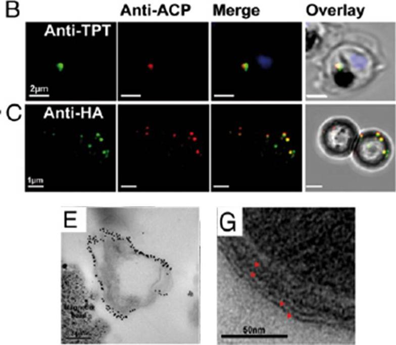
Purity and integrity of isolated apicoplasts. (A) Protein samples from each purification step were examined by. (B) Immunofluorescence of intracellular trophozoite showing apicoplast with apicoplast stromal marker ACP (red), apicoplast outer membrane marker PfoTPT (green), and parasite nucleus (blue). (C) Purified apicoplasts bound to magnetic beads seen via anti-ACP (red) and anti-HA tagged TPT (green) labeling. (D–G) Electron micrographs showing purified apicoplasts in vicinity of magnetic beads (E), as confirmed by anti-PfoTPT immunogold labeling (E), four surrounding membranes (G), and ribosome-like particles within (G).Botté CY, Yamaryo-Botté Y, Rupasinghe TW, Mullin KA, MacRae JI, Spurck TP, Kalanon M, Shears MJ, Coppel RL, Crellin PK, Maréchal E, McConville MJ, McFadden GI. Atypical lipid composition in the purified relict plastid (apicoplast) of malaria parasites. Proc Natl Acad Sci U S A. 2013 Apr 110(18):7506-11
See original on MMP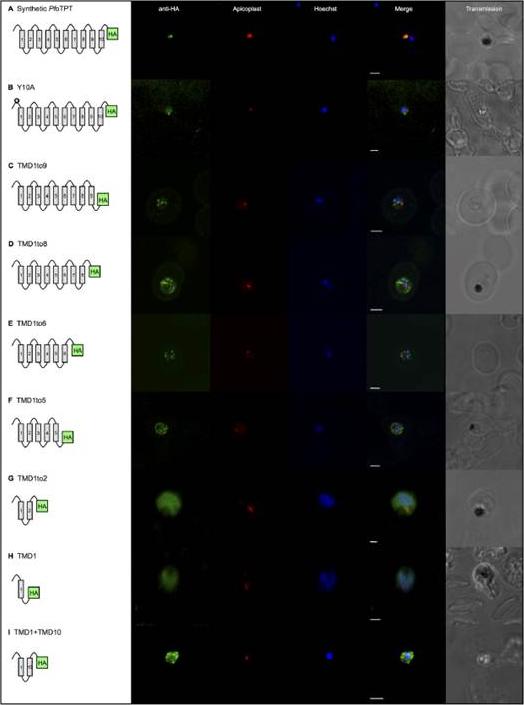
Gene expression constructs defining elements of PfoTPT essential for targeting to the outer membrane of the apicoplast. TMDs are represented by numbered boxes and are joined by loops. All PfoTPT constructs are episomally expressed under the PfCRT promoter and tagged with triple HA at the C-terminus detected with anti-HA and secondary antibody conjugated to FITC (green) in parasites within erythrocytes. Co-localisation of the apicoplast using antisera against apicoplast stromal marker, ACP, is shown in red. Nuclei are stained with Hoechst (blue), and transmitted light images of the parasites within their host red blood cell are shown on the right. Scale bars = 2μm. A. Full length, synthetic PfoTPT colocalises with ACP. B. Point mutation of tyrosine residue at position 10 (✪) in the synthetic PfoTPT (Y10A) abrogates targeting to the apicoplast showing no co-localisation with the apicoplast marker. C. Removal of TMD 10 (TMD1to9) abrogates targeting to the apicoplast showing no co-localisation with the apicoplast marker. D. Removal of TMDs 9 and 10 (TMD1to8) abrogates targeting to the apicoplast showing no co-localisation with the apicoplast marker. E. Removal of TMDs 7, 8, 9 and 10 (TMD1to6) abrogates targeting to the apicoplast showing no co-localisation with the apicoplast marker. F. Removal of TMDs 6, 7, 8, 9 and 10 (TMD1to5) abrogates targeting to the apicoplast showing no co-localisation with the apicoplast marker. G. Removal of TMDs 3, 4, 5, 6, 7, 8, 9 and 10 (TMD1to2) abrogates targeting to the apicoplast showing no co-localisation with the apicoplast marker and diffuse staining throughout the parasite. H. Removal of TMDs 2, 3, 4, 5, 6, 7, 8, 9 and 10 (TMD1) abrogates targeting to the apicoplast showing no co-localisation with the apicoplast marker and diffuse staining throughout the parasite. I. A combination of TMD1 and TMD10 (TMD1+TMD10) was not sufficient to reconstitute targeting to the apicoplast, showing no co-localisation with the apicoplast markerLim L, Sayers CP, Goodman CD, McFadden GI. Targeting of a Transporter to the Outer Apicoplast Membrane in the Human Malaria Parasite Plasmodium falciparum. PLoS One. 2016 11(7):e0159603.
See original on MMP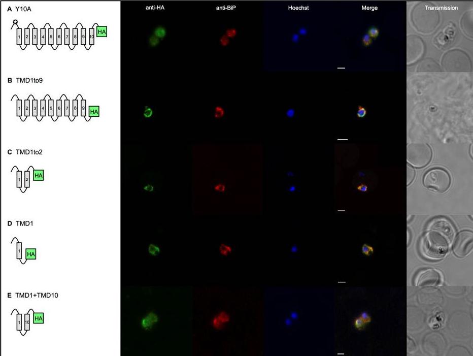
Select gene expression constructs not targeted to the apicoplast are now co-localised with the endoplasmic reticulum. TMDs are represented by boxes and are joined by loops. All PfoTPT constructs are episomally expressed under the PfCRT promoter and tagged with triple HA at the C-terminus detected with anti-HA and secondary antibody conjugated to FITC (green). The parasite ER is detected with anti-BiP (red). Nuclei are stained with Hoechst (blue), and transmitted light images of the parasites within their host red blood cell are shown at right. Scale bars = 2μm. A. Point mutation of tyrosine residue at position 10 (✪) in the synthetic PfoTPT (Y10A) relocates protein from the apicoplast (B) to the ER, as well as the plasma. B. Removal of TMD 10 (TMD1to9), which completely abrogates targeting to the apicoplast (C), results in perinuclear localisation of the protein that almost entirely overlaps with the ER marker, BiP. C. Removal of TMDs 3, 4, 5, 6, 7, 8, 9 and 10 (TMD1to2), which completely abrogates targeting to the apicoplast (G), results in re-localisation of most of the protein to the ER. D. Removal of TMDs 2, 3, 4, 5, 6, 7, 8, 9 and 10 (TMD1), which completely abrogates targeting to the apicoplast (H), primarily results in localisation of most of the protein to the ER. E. Recombining TMD1 with TMD10 (TMD1+10) was unable to reconstitute apicoplast targeting (I) and resulted in some perinuclear ER targeting, plus some parasite plasma membrane targeting.Lim L, Sayers CP, Goodman CD, McFadden GI. Targeting of a Transporter to the Outer Apicoplast Membrane in the Human Malaria Parasite Plasmodium falciparum. PLoS One. 2016 11(7):e0159603.
See original on MMP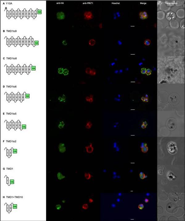
Gene expression constructs not targeted to the apicoplast now co-localised with the plasma membrane. TMDs are represented by boxes and are joined by loops. All PfoTPT constructs are episomally expressed under the PfCRT promoter and tagged with triple HA at the C-terminus detected with anti-HA and secondary antibody conjugated to FITC (green). The parasite plasma membrane is detected with anti-PfNTP1 (red). Nuclei are stained with Hoechst (blue), and transmitted light images of the parasites within their host red blood cell are shown at right. Scale bars = 2μm. A. Point mutation of tyrosine residue at position 10 (✪) in the synthetic PfoTPT (Y10A) relocates protein from the apicoplast (B) to the plasma membrane. B. Removal of TMD 10 (TMD1to9), which completely abrogates targeting to the apicoplast (C), does not result in plasma membrane localisation. C. Removal of TMDs 9 and 10 (TMD1to8), which completely abrogates targeting to the apicoplast (D), results in some protein localisation to the plasma membrane. D. Removal of TMDs 7, 8, 9 and 10 (TMD1to6), which completely abrogates targeting to the apicoplast (E), results in localisation of the protein that mostly overlaps with the parasite plasma membrane marker, PfNT1. Note that the erythrocyte is infected with three separate parasites. E. Removal of TMDs 6, 7, 8, 9 and 10 (TMD1to5), which completely abrogates targeting to the apicoplast (F), results in localisation of the protein that mostly overlaps with the parasite plasma membrane marker, PfNT1. Note that the erythrocyte is infected with two separate parasites. F. Removal of TMDs 3, 4, 5, 6, 7, 8, 9 and 10 (TMD1to2), which completely abrogates targeting to the apicoplast (G), results in some re-localisation of the protein to the plasma membrane. G. Removal of TMDs 2, 3, 4, 5, 6, 7, 8, 9 and 10 (TMD1), which completely abrogates targeting to the apicoplast (H), results in some localisation of the protein to the plasma membrane. H. Recombining TMD1 with TMD10 (TMD1+10) was unable to reconstitute apicoplast targeting (I) and resulted in some plasma membrane targeting.Lim L, Sayers CP, Goodman CD, McFadden GI. Targeting of a Transporter to the Outer Apicoplast Membrane in the Human Malaria Parasite Plasmodium falciparum. PLoS One. 2016 11(7):e0159603.
See original on MMP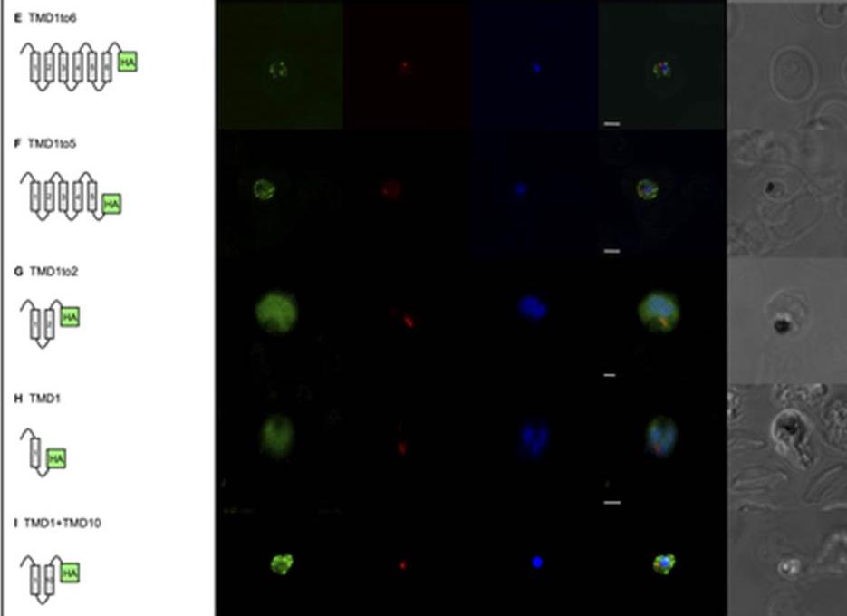
Gene expression constructs not targeted to the apicoplast now co-localised with the plasma membrane. Removal of TMDs 7, 8, 9 and 10 (TMD1to6), which completely abrogates targeting to the apicoplast (Fig 2E), results in localisation of the protein that mostly overlaps with the parasite plasma membrane marker, PfNT1. Note that the erythrocyte is infected with three separate parasites. E. Removal of TMDs 6, 7, 8, 9 and 10 (TMD1to5), which completely abrogates targeting to the apicoplast (Fig 2F), results in localisation of the protein that mostly overlaps with the parasite plasma membrane marker, PfNT1. Note that the erythrocyte is infected with two separate parasites. F. Removal of TMDs 3, 4, 5, 6, 7, 8, 9 and 10 (TMD1to2), which completely abrogates targeting to the apicoplast (Fig 2G), results in some re-localisation of the protein to the plasma membrane. G. Removal of TMDs 2, 3, 4, 5, 6, 7, 8, 9 and 10 (TMD1), which completely abrogates targeting to the apicoplast (Fig 2H), results in some localisation of the protein to the plasma membrane. H. Recombining TMD1 with TMD10 (TMD1+10) was unable to reconstitute apicoplast targeting (Fig 2I) and resulted in some plasma membrane targeting.Lim L, Sayers CP, Goodman CD, McFadden GI. Targeting of a Transporter to the Outer Apicoplast Membrane in the Human Malaria Parasite Plasmodium falciparum. PLoS One. 2016 11(7):e0159603.
See original on MMP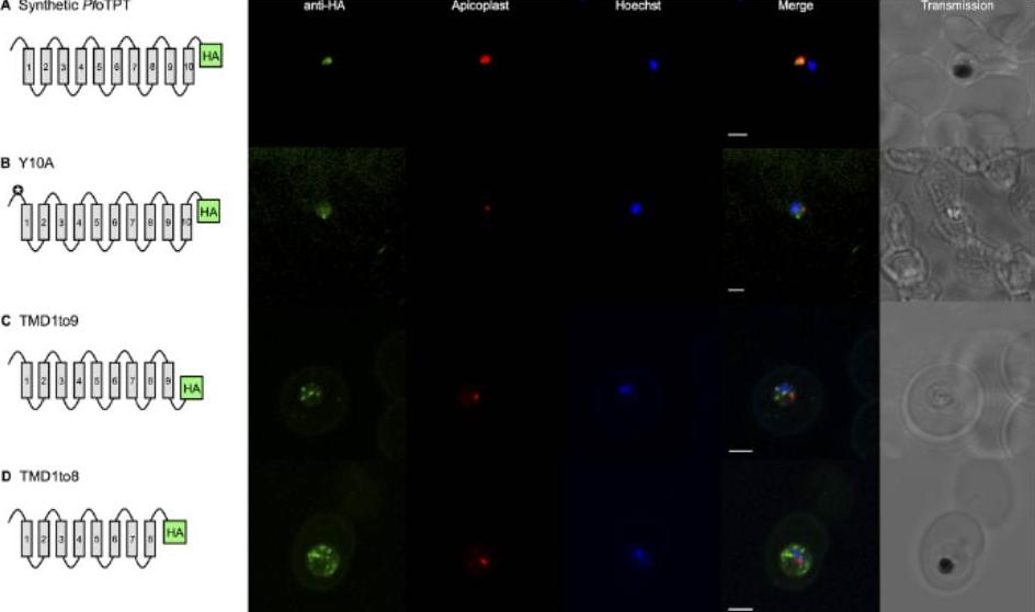
Gene expression constructs defining elements of PfoTPT essential for targeting to the outer membrane of the apicoplast. TMDs are represented by numbered boxes and are joined by loops. All PfoTPT constructs are episomally expressed under the PfCRT promoter and tagged with triple HA at the C-terminus detected with anti-HA and secondary antibody conjugated to FITC (green) in parasites within erythrocytes. Co-localisation of the apicoplast using antisera against apicoplast stromal marker, ACP, is shown in red. Nuclei are stained with Hoechst (blue), and transmitted light images of the parasites within their host red blood cell are shown on the right. Scale bars = 2μm. A. Full length, synthetic PfoTPT colocaliseswith ACP. B. Point mutation of tyrosine residue at position 10 (✪) in the synthetic PfoTPT (Y10A) abrogates targeting to the apicoplast showing no co-localisation with the apicoplast marker. C. Removal of TMD 10 (TMD1to9) abrogates targeting to the apicoplast showing no co-localisation with the apicoplast marker. D. Removal of TMDs 9 and 10 (TMD1to8) abrogates targeting to the apicoplast showing no co-localisation with the apicoplast marker. marker.Lim L, Sayers CP, Goodman CD, McFadden GI. Targeting of a Transporter to the Outer Apicoplast Membrane in the Human Malaria Parasite Plasmodium falciparum. PLoS One. 2016 11(7):e0159603.
See original on MMPMore information
| PlasmoDB | PY17X_1109000 |
| GeneDB | PY17X_1109000 |
| Malaria Metabolic Pathways | Localisation images Pathways mapped to |
| Previous ID(s) | null |
| Orthologs | PBANKA_1107900 , PCHAS_1107600 , PF3D7_0508300 , PKNH_1025300 , PVP01_1025700 , PVX_097975 |
| Google Scholar | Search for all mentions of this gene |