PY17X_1004300 ag-1 blood stage membrane protein homologue
Disruptability [+]
| Species | Disruptability | Reference | Submitter | |
|---|---|---|---|---|
| P. berghei ANKA |
Refractory |
PlasmoGEM (Barseq) | PlasmoGEM | |
| P. falciparum 3D7 |
Refractory |
22986493 | Theo Sanderson, Wellcome Trust Sanger Institute | |
| P. falciparum 3D7 |
Refractory |
biorxiv.org/content/10.1101/2020.11.26.400549v2 | Benjamin Liffner, The University of Adelaide | |
Mutant phenotypes [+]
| Species | Stage | Phenotype | Reference | Submitter |
|---|---|---|---|---|
| P. falciparum 3D7 | Asexual |
Invasion defect |
https://www.biorxiv.org/content/10.1101/2020.11.26.400549v2
(Knock down)
Aberrantly elongated rhoptries |
Benjamin Liffner, The University of Adelaide |
Imaging data (from Malaria Metabolic Pathways)
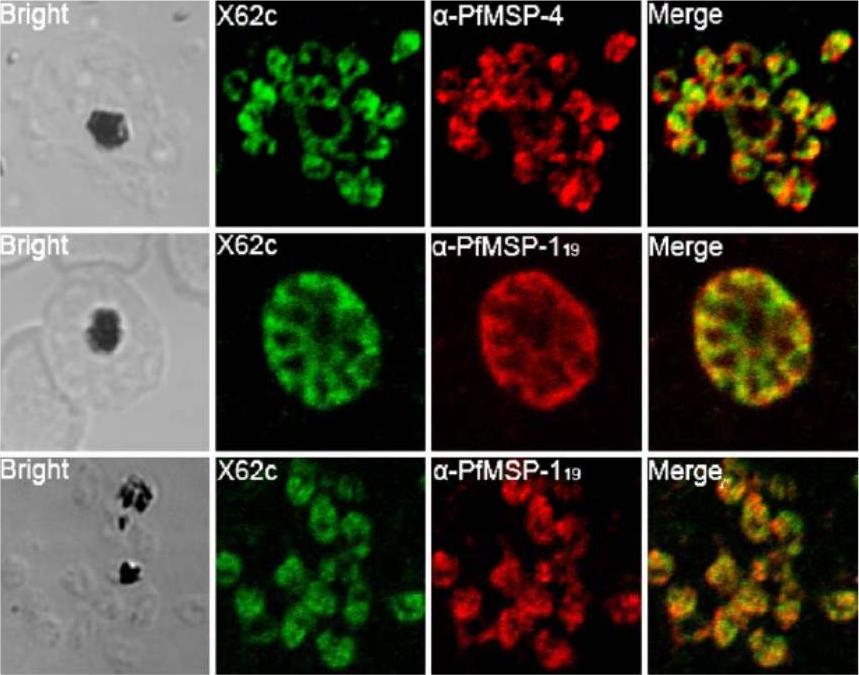
Co-localization of PfMAg-1 with PfMSP-4 or PfMSP-119 at late schizont/merozoite parasites examined by confocal fluorescent microscopy. PfMSP-119 is the 19-kDa C-terminal fragment of PfMSP-1 that remains membrane-bound on the invading merozoite Top: Merozoite detected with antibodies X62c (against PfMAg-1) and α-PfMSP-4. Middle: Mature schizont detected with antibodies X62c and α-PfMSP-119. Bottom: Merozoite detected with antibodies X62c and α-PfMSP-119. Bright field images are shown on the left panel. PfMSP-1 and PfMSP-4 are two of the major merozoite surface proteins characterized. Schizonts/merozoite of 3D7 strain, PfMAg-1 co-localized very well with PfMSP-119, and partially with PfMSP-4, in the typical bunch-of-grapes fluorescence pattern haracteristic of merozoite surface proteins.Gao YH, Li HL, Lu Y, Gao FM, Lin YH, Zhou HC, Zhang LH, Wang H. Identification of a vaccine candidate antigen, PfMAg-1, from Plasmodium falciparum with monoclonal antibody M26-32. Parasitol Res. 2009 105:1723-32. PMID: 19777263
See original on MMP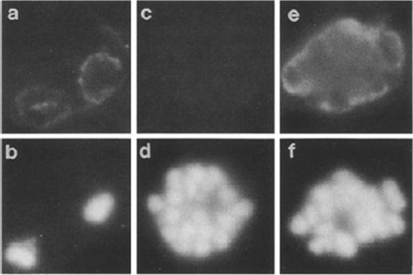
Localization of Ag1, showing dual immunofluorescence staining of Ag1 with human monospecific antibodies against Ag1 or control serum, a Immunofluorescence of trophozoites with human anti-Ag1 antibodies, b DNA staining of trophozoites, c Immunofluorescence with control serum, d DNA staining of schizonts, e Immunofluorescence of schizonts with human anti-Ag1 antibodies, f DNA staining of schizonts. Antibodies showed clear reactivity with trophozoite- and schizont-infected erythrocytes localized on the surface of the parasites. Neither individual merozoites within the schizont nor free merozoites were labelled.Jakobsen PH, Jepsen S, Riley EM, Theander TG, Grellier P, Lihme A, Hviid L, Dziegiel M, Schrevel J. Biochemical characterization, localization and immunostimulating properties of a soluble glycoprotein, Ag1, isolated from in vitro cultures of Plasmodium falciparum. Parasitol Res. 1990;76(8):657-61.
See original on MMP
Confocal images of FITC PfMAg-1 in mature stage P. falciparum 3D7 infected erythrocytes examined by indirect immunofluorescent assay. Trophozoite stage parasites were immunostained with McAb X62c followed by FITC-labeledsecondary antibody. In addition to the fluorescent labeling on the bodies of trophozoite parasites, dispersed fluorescence patches were observed underneath the parasite-infected erythrocyte membrane (a), while no fluorescence was observed when normal mouse serum was used (b)Co-localization of PfMAg-1 with PfMSP-4 or PfMSP-119 at late schizont/merozoite parasites examined by confocal fluorescent microscopy. a Merozoite detected with antibodies X62c and α-PfMSP-4. b Mature schizont detected with antibodies X62c and α-PfMSP-119. c Merozoite detected with antibodies X62c and α-PfMSP-119. Bright field images are shown on the left panel.Gao YH, Li HL, Lu Y, Gao FM, Lin YH, Zhou HC, Zhang LH, Wang H. Identification of a vaccine candidate antigen, PfMAg-1, from Plasmodium falciparum with monoclonal antibody M26-32. Parasitol Res. 2009 105(6):1723-32.
See original on MMPMore information
| PlasmoDB | PY17X_1004300 |
| GeneDB | PY17X_1004300 |
| Malaria Metabolic Pathways | Localisation images Pathways mapped to |
| Previous ID(s) | null |
| Orthologs | PBANKA_1002900 , PCHAS_1003800 , PF3D7_0405200 , PKNH_0303300 , PVP01_0304600 , PVX_000980 |
| Google Scholar | Search for all mentions of this gene |