PY17X_0713100 high mobility group protein B2 (HMGB2)
Disruptability [+]
| Species | Disruptability | Reference | Submitter | |
|---|---|---|---|---|
| P. yoelii yoelii 17X |
Possible |
RMgm-162 | Imported from RMgmDB | |
| P. berghei ANKA |
Possible |
RMgm-1251 | Imported from RMgmDB | |
| P. berghei ANKA |
Possible |
RMgm-4831 | Imported from RMgmDB | |
| P. falciparum 3D7 |
Possible |
36506024 Functional characterization of PfHMGB2 by gene deletion (Pfhmgb2¯) showed that knockout parasites develop normally as asexual stages and undergo gametocytogenesis. Transmission experiments revealed that Pfhmgb2¯ can infect mosquitoes and develop as oocyst stages. However, transmission was reduced compared to wild type (WT) parasites and as a consequence, the salivary gland sporozoites were reduced in number. |
Theo Sanderson, Francis Crick Institute | |
Mutant phenotypes [+]
| Species | Stage | Phenotype | Reference | Submitter |
|---|---|---|---|---|
| P. yoelii yoelii 17X | Asexual |
Difference from wild-type |
RMgm-162
A slight growth delay of the asexual blood stages, resulting in a 'delay in the onset of parasitemia'. |
Imported from RMgmDB |
| P. yoelii yoelii 17X | Gametocyte |
No difference |
RMgm-162
Normal gametocyte production (as determined by counting mature gametocytes in Giemsa stained slides). Normal exflagellation of male gametocytes. |
Imported from RMgmDB |
| P. yoelii yoelii 17X | Ookinete |
Difference from wild-type |
RMgm-162
Reduction of in vitro ookinete production (52-61% compared to wild type). |
Imported from RMgmDB |
| P. yoelii yoelii 17X | Oocyst |
Difference from wild-type |
RMgm-162
the mean number of oocysts in mutant infected mosquito was ~10% of the mean oocyst number seen in wild type infected mosquitoes. The mutant oocysts produced viable sporozoites that were infectious to mice. |
Imported from RMgmDB |
| P. yoelii yoelii 17X | Sporozoite |
No difference |
RMgm-162 | Imported from RMgmDB |
| P. yoelii yoelii 17X | Liver |
No difference |
RMgm-162 | Imported from RMgmDB |
| P. berghei ANKA | Asexual |
Difference from wild-type |
RMgm-1251
Evidence is presented for normal growth of blood stages but reduction of cerebral complications in C57Bl6 mice that are susceptible to ECM (Experimental Cerebral Malaria). |
Imported from RMgmDB |
| P. berghei ANKA | Asexual |
Difference from wild-type |
RMgm-4831
Reduced growth of blood stages and C57Bl/6 are able to clear the infection after day 15 (in contrast to wild type infections where mice die from hyperparasitemia) |
Imported from RMgmDB |
| P. falciparum 3D7 | Asexual |
No difference |
36506024 Functional characterization of PfHMGB2 by gene deletion (Pfhmgb2¯) showed that knockout parasites develop normally as asexual stages and undergo gametocytogenesis. Transmission experiments revealed that Pfhmgb2¯ can infect mosquitoes and develop as oocyst stages. However, transmission was reduced compared to wild type (WT) parasites and as a consequence, the salivary gland sporozoites were reduced in number. |
Theo Sanderson, Francis Crick Institute |
| P. falciparum 3D7 | Oocyst |
Difference from wild-type |
36506024 Functional characterization of PfHMGB2 by gene deletion (Pfhmgb2¯) showed that knockout parasites develop normally as asexual stages and undergo gametocytogenesis. Transmission experiments revealed that Pfhmgb2¯ can infect mosquitoes and develop as oocyst stages. However, transmission was reduced compared to wild type (WT) parasites and as a consequence, the salivary gland sporozoites were reduced in number. |
Theo Sanderson, Francis Crick Institute |
Imaging data (from Malaria Metabolic Pathways)
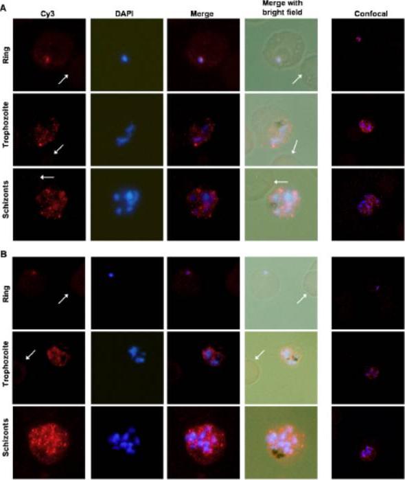
Localization of PfHMGB1 and PfHMGB2 in 3D7 Plasmodium falciparum parasite. mmunofluorescence in ring, trophozoite and schizonts of P. falciparum with anti-HMGB1 [A] and anti-HMGB2 [B] using IFA and confocal microscopy were analyzed. In column I, staining with anti-HMGBs is shown, while in column II staining with DAPI, column III merge of staining with anti-HMGBs and DAPI, column IV, merge of staining by anti-HMGBs and DAPI with the bright field of the corresponding parasites is presented. Column V, confocal images are shown. Uninfected RBCs were represented with arrowhead, where no fluorescence signal was observed. Both gene products were present in the nucleus at the ring stage . However,significant amount of these proteins were also present inthe parasite cytosol at the trophozoite and schizont stagesKumar K, Singal A, Rizvi MM, Chauhan VS. High mobility group box (HMGB) proteins of Plasmodium falciparum: DNA binding proteins with pro-inflammatory activity. Parasitol Int. 2008 57(2):150-7.
See original on MMP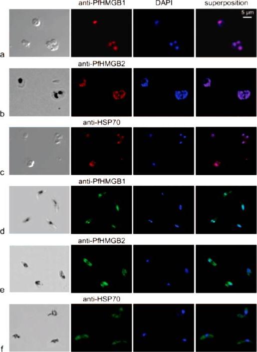
Immunofluorescence localization of PfHMGB1 and PfHMGB2 in asexual (a, b, and c) and sexual (d, e, and f) stages of Plasmodium erythrocytic development. Paraformaldehyde-fixed parasites were labeled with mouse anti-PfHMGB1 and anti-PfHMGB2 antibodies (1:200) and FITC-conjugated anti-mouse IgG (1:100); DNA was stained with DAPI (1:100). Merged fluorescent signals are shown in the “superposition” column. Cells were visualized by phase-contrast (a and c) or transmission (b, d, e, and f) microscopy. Panels: a, trophozoites; b, trophozoite and schizont; c, trophozoites; d to f, gametocytes. Anti-PfHMGB and anti-HSP70 fluorescence is red for panels a to c and green for panels d to f. We also compared the localizations of both factors in asexual (red immunofluorescence) and gametocyte (green immunofluorescence) stages. As already mentioned, the two PfHMGB factors (lanes a and b) appeared to be located mainly in the nucleus of the asexual stages (rings, trophozoites, and schizonts), whereas the HSP70 protein (lane c) was also found in the parasite cytoplasm. Surprisingly, in addition to its nuclear localization, PfHMGB2 could also be readily detected within the cytoplasm of different stages (IV and V) of gametocytes (lanes e), as also observed for the HSP70 protein (lane f), whereas PfHMGB1 was associated mainly with the nucleus of gametocytes, as in asexual parasites (lanes d and a).Briquet S, Boschet C, Gissot M, Tissandié E, Sevilla E, Franetich JF, Thiery I, Hamid Z, Bourgouin C, Vaquero C. High-mobility-group box nuclear factors of Plasmodium falciparum. Eukaryot Cell. 2006 5(4):672-82.
See original on MMP
Localization of PfHMGB1 and PfHMGB2 in 3D7 Plasmodium falciparum parasite. mmunofluorescence in ring, trophozoite and schizonts of P. falciparum with anti-HMGB1 [A] and anti-HMGB2 [B] using IFA and confocal microscopy were analyzed. In column I, staining with anti-HMGBs is shown, while in column II staining with DAPI, column III merge of staining with anti-HMGBs and DAPI, column IV, merge of staining by anti-HMGBs and DAPI with the bright field of the corresponding parasites is presented. Column V, confocal images are shown. Uninfected RBCs were represented with arrowhead, where no fluorescence signal was observed. Both gene products were present in the nucleus at the ring stage . However,significant amount of these proteins were also present inthe parasite cytosol at the trophozoite and schizont stagesKumar K, Singal A, Rizvi MM, Chauhan VS. High mobility group box (HMGB) proteins of Plasmodium falciparum: DNA binding proteins with pro-inflammatory activity. Parasitol Int. 2008 57(2):150-7.
See original on MMPMore information
| PlasmoDB | PY17X_0713100 |
| GeneDB | PY17X_0713100 |
| Malaria Metabolic Pathways | Localisation images Pathways mapped to |
| Previous ID(s) | null |
| Orthologs | PBANKA_0712900 , PCHAS_0722000 , PF3D7_0817900 , PVP01_0517400 , PVX_089520 |
| Google Scholar | Search for all mentions of this gene |