PVX_122810 hypothetical protein, conserved
Disruptability [+]
| Species | Disruptability | Reference | Submitter | |
|---|---|---|---|---|
| P. berghei ANKA |
Refractory |
RMgm-216 | Imported from RMgmDB | |
| P. falciparum 3D7 |
Refractory |
29579782 Several attempts using SLI |
Theo Sanderson, Wellcome Trust Sanger Institute | |
| P. falciparum 3D7 |
Possible |
USF piggyBac screen (Insert. mut.) | USF PiggyBac Screen | |
Mutant phenotypes [+]
| Species | Stage | Phenotype | Reference | Submitter |
|---|---|---|---|---|
| P. falciparum 3D7 | Asexual |
Attenuated |
29579782 (Knock down)
glmS-based knockdown produced "A mild reduction in parasitaemia of around 30 % after three cycles" |
Theo Sanderson, Wellcome Trust Sanger Institute |
Imaging data (from Malaria Metabolic Pathways)
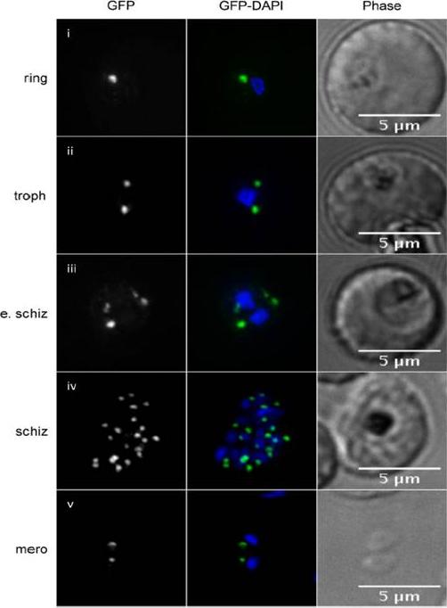
PfPRP2-RFA is constitutively expressed throughout the erythrocytic cycle. Dynamics of the localization of PfPRP2-RFA throughout the asexual stages. In ring stage parasites, a single dot offluorescence is seen close to the nucleus In trophozoites, the dot starts to multiply, prior to nuclear division. Multiplication of the PfPRP2-RFA signal continues once nuclear division is undertaken . In free merozoites, each parasite inherits one locus of PfPRP2-RFA fluorescence . Thefluorescence of PfPRP2-RFA is pseudo-colored in green and the nuclei of parasites were stained with DAPI (blue). Scale bar represents 5μm. Troph: trophozoite; E. schiz: early schizont; Schiz: schizont; Mero: merozoite.Hallée S, Richard D. Evidence that the Malaria Parasite Plasmodium falciparum Putative Rhoptry Protein 2 Localizes to the Golgi Apparatus throughout the Erythrocytic Cycle. PLoS One. 2015 Sep 16;10(9):e0138626. PMID:
See original on MMP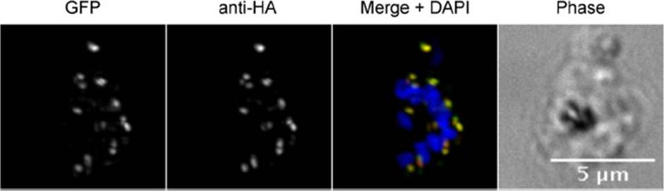
Generation of a PfPRP2-RFA tagged parasite line. IFA using anti-HA (red) or epifluorescence of the GFP (green) on the PfPRP2-RFA tagged line shows an identical punctate pattern. Scale bar represents 5 μm. Nuclei of parasites were stain with DAPI (blue). A typical punctate pattern consistent with the proposed apical tip localization was observed for both the anti-HA antibody and the native GFP fluorescence, in line with PfPRP2 being a potential rhoptry protein.Hallée S, Richard D. Evidence that the Malaria Parasite Plasmodium falciparum Putative Rhoptry Protein 2 Localizes to the Golgi Apparatus throughout the Erythrocytic Cycle. PLoS One. 2015 Sep 16;10(9):e0138626.
See original on MMP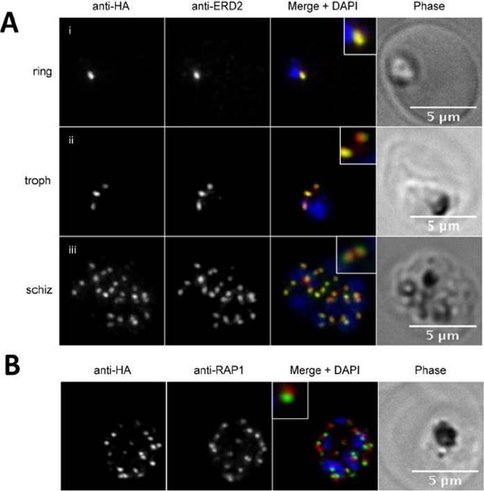
PfPRP2-RFA overlaps extensively with the Golgi apparatus throughout the erythrocytic cycle. (A) In ring stages, PfPRP2-RFA is found as a single punctate pattern colocalizing with the cis-Golgi marker ERD2 (Ai). In trophozoites, prior to nuclear division, the pattern associated with both PfPRP2-RFA and ERD2 becomes triple dots, showing the division of the Golgi, prior to nuclear replication (Aii). In schizont stages, themultiple PfPRP2-RFA signals still colocalize with the cis-Golgi protein ERD2 (Aiii). (B) In mature schizonts, PfPRP2-RFA is found in close proximity but does not overlap with the rhoptry marker RAP1. RAP1, rhoptry associated protein 1. Nuclei of parasites were stained with DAPI (blue). The fluorescence of PfPRP2-RFA is pseudocolored in green and other markers are in red. Scale bar represents 5μm.Hallée S, Richard D. Evidence that the Malaria Parasite Plasmodium falciparum Putative Rhoptry Protein 2 Localizes to the Golgi Apparatus throughout the Erythrocytic Cycle. PLoS One. 2015 Sep 16;10(9):e0138626.
See original on MMPMore information
| PlasmoDB | PVX_122810 |
| GeneDB | PVX_122810 |
| Malaria Metabolic Pathways | Localisation images Pathways mapped to |
| Previous ID(s) | Pv122810 |
| Orthologs | PBANKA_1418300 , PCHAS_1420100 , PF3D7_1320000 , PKNH_1420600 , PVP01_1420800 , PY17X_1420000 |
| Google Scholar | Search for all mentions of this gene |