PVX_119490 ATP-dependent Clp protease proteolytic subunit, putative
Disruptability [+]
| Species | Disruptability | Reference | Submitter | |
|---|---|---|---|---|
| P. falciparum 3D7 |
Refractory |
USF piggyBac screen (Insert. mut.) | USF PiggyBac Screen | |
Mutant phenotypes [+]
| Species | Stage | Phenotype | Reference | Submitter |
|---|---|---|---|---|
| P. falciparum 3D7 | Asexual | !!
Not tested |
http://biorxiv.org/content/biorxiv/early/2017/06/20/080408.full.pdf
(Conditional)
No difference was seen on KD with glucosamine but since KD is partial it is difficult to conclude dispensability |
Theo Sanderson, Wellcome Trust Sanger Institute |
Imaging data (from Malaria Metabolic Pathways)
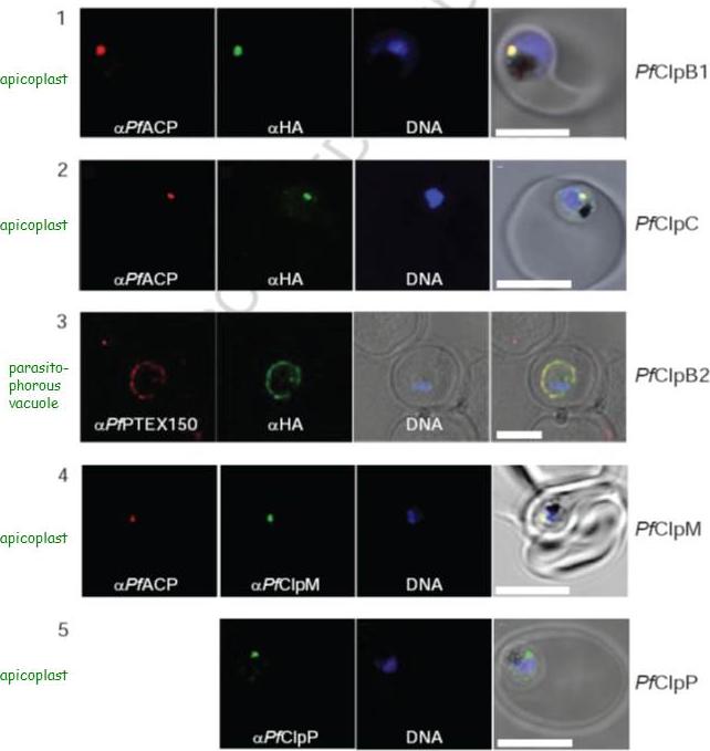
Localization of PfClpB1, PfClpC, PfClpB2, PfClpM, and PfClpP of P. falciparum was tested by immunofluorescent colocalization. In panels 1 and 2, PfClpB1-Strp-3×HA or PfClpC-Strp-3×HA parasites were probed with the apicoplast marker anti-ACP PFB0385w antisera (red), with anti-HA antisera (green), and with Hoescht 33342 to stain for DNA (blue). In panel 3, PfClpB2-Strp-3×HA parasites were probed with the parasitophorous vacuole marker anti-PTEX150 antisera (red), with anti-HA antisera (green), and with Hoescht 33342 (blue). In panels 4 and 5, wild type 3D7 strain was probed with anti-ACP antisera (red), with anti-PfClpM antisera or anti-ClpP antisera (green), and with Hoescht 33342 (blue). For all panels, the right most image represents the merged fluorescence images with the DIC or transmission image. The scale bars correspond to 5 mm.El Bakkouri M, Pow A, Mulichak A, Cheung KL, Artz JD, Amani M, Fell S, de Koning T, Goodman CD, McFadden GI, Ortega J, Hui R, Houry WA. The Clp chaperones and proteases of the human malaria parasite Plasmodium falciparum. J Mol Biol. 2010 404(3):456-77
See original on MMP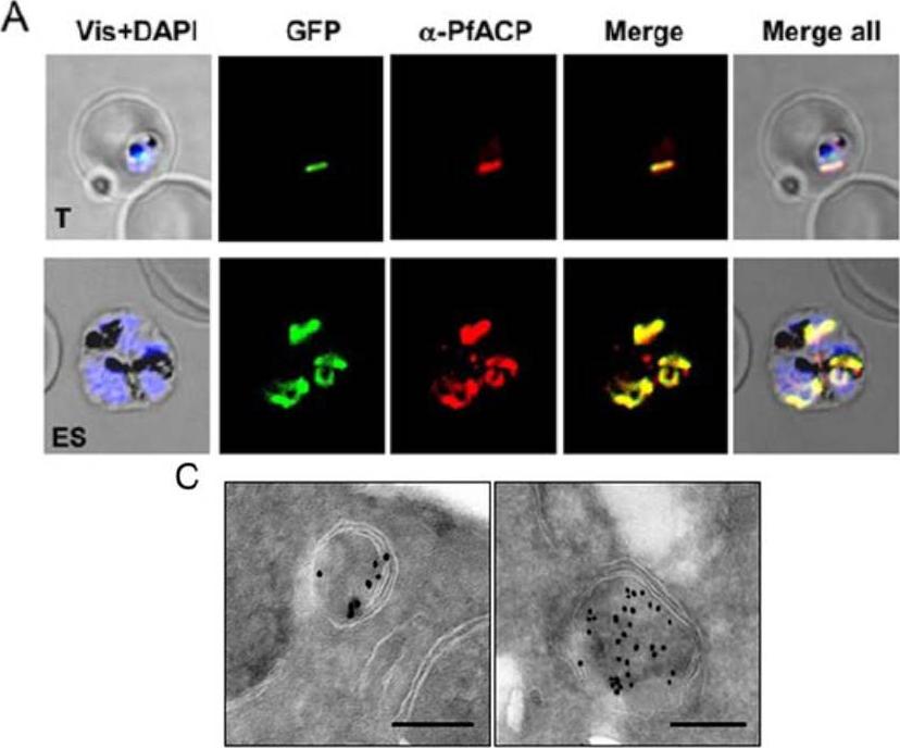
Immunofluorescence assay and immuno-electron microscopy to localize PfClpP. (A) Transgenic parasites expressing PfClpP-N-GFP were immuno-stained with antibodies specific to the apicoplast localized acyl carrier protein (ACP PFB0385w) (red). The parasite nuclei were stained with DAPI (blue) and slides were visualized by confocal laser scanning microscope. The PfClpP-N-GFP fusion protein and ACP were colocalized in the parasite apicoplast. (C) Localization of PfClpP by immuno-electron microscopy. Ultra thin sections of transgenic P. falciparum parasites expressing PfClpP-N-GFP were labeled with anti-GFP antibody and gold labeled secondary antibody. Labeling was observed in the apicoplast having characteristic four membranes. Scale bar = 250nm. Rathore S, Sinha D, Asad M, Böttcher T, Afreen F, Chauhan VS, Gupta D, Sieber S, Mohmmed A. A cyanobacterial serine protease of Plasmodium falciparum is targeted to the apicoplast and plays important role in its growth and development. Mol Microbiol. 2010 ) 77: 873–890.
See original on MMP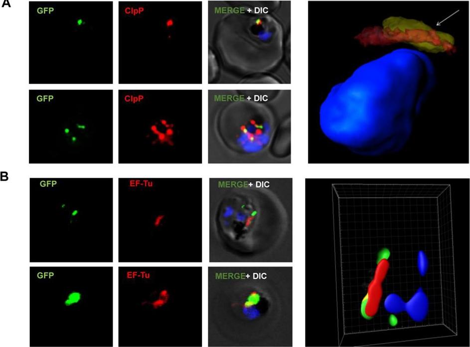
Confocal microscopy images of transgenic parasites PfOTU-RFA (green) and immune-stained for apicoplast targeted protein ClpP (A) and apicoplast resident protein EF-Tu (B). For each set, a three-dimensional reconstruction was developed using series of Z-stacks from confocal images with Imaris software. ClpP and EF-Tu staining are shown in red; the ClpP/EF-Tu and GFP co-localising area is seen as yellow staining. In all the cases the PfOTU-GFP labelled vesicles were observed to be in close proximity or partially overlapping with apicoplast staining. The 3D image reconstruction based upon confocal Z-stacks showed these vesicles to merge into the apicoplast in some cases.Datta G, Hossain ME, Asad M, Rathore S, Mohmmed A. Plasmodium falciparum OTU-like cysteine protease (PfOTU) is essential for apicoplast homeostasis and associates with non-canonical role of Atg8. Cell Microbiol. 2017 Apr 18. [Epub ahead of print]
See original on MMPMore information
| PlasmoDB | PVX_119490 |
| GeneDB | PVX_119490 |
| Malaria Metabolic Pathways | Localisation images Pathways mapped to |
| Previous ID(s) | Pv119490 |
| Orthologs | PBANKA_0405700 , PCHAS_0406600 , PF3D7_0307400 , PKNH_0835300 , PVP01_0832800 , PY17X_0408000 |
| Google Scholar | Search for all mentions of this gene |