PVX_113890 enoyl-acyl carrier protein reductase
Disruptability [+]
| Species | Disruptability | Reference | Submitter | |
|---|---|---|---|---|
| P. berghei ANKA |
Possible |
RMgm-256 | Imported from RMgmDB | |
| P. berghei ANKA |
Possible |
RMgm-197 | Imported from RMgmDB | |
| P. falciparum 3D7 |
Possible |
19068099 | Theo Sanderson, Wellcome Trust Sanger Institute | |
Mutant phenotypes [+]
| Species | Stage | Phenotype | Reference | Submitter |
|---|---|---|---|---|
| P. berghei ANKA | Asexual |
No difference |
RMgm-256 | Imported from RMgmDB |
| P. berghei ANKA | Asexual |
No difference |
RMgm-197 | Imported from RMgmDB |
| P. falciparum 3D7 | Asexual |
No difference |
19068099 | Theo Sanderson, Wellcome Trust Sanger Institute |
| P. berghei ANKA | Gametocyte |
No difference |
RMgm-197 | Imported from RMgmDB |
| P. berghei ANKA | Ookinete |
No difference |
RMgm-197 | Imported from RMgmDB |
| P. berghei ANKA | Oocyst |
No difference |
RMgm-197 | Imported from RMgmDB |
| P. berghei ANKA | Sporozoite |
Difference from wild-type |
RMgm-197
Normal numbers of salivary gland sporozoites are formed. Sporozoites have a strongly reduced infectivity to mice as shown by a significant delay of blood infections after intravenous inoculation of sporozoites. |
Imported from RMgmDB |
| P. berghei ANKA | Liver |
Difference from wild-type |
RMgm-197
Normal numbers of salivary gland sporozoites are formed. Sporozoites have a strongly reduced infectivity to mice as shown by a significant delay of blood infections after intravenous inoculation of sporozoites. Sporozoites showed normal cell traversal, hepatocyte invasion rates and initial stages of intrahepatic development. The formation of merozoites within the liver schizonts was strongly affected. Abnormal progression of nuclear division became apparent. 99.5% of the schizonts was MSP1 negative. No mature merozoites were formed and released.Injection of 1000 PbfabI SPZs produced a blood-stage infection in only 5/16 mice, with the infected mice showing a delay in patency of 4 days. Increasing the PbfabI inoculum to 10,000 SPZs resulted in patent infections in 13/15 mice, with those mice again showing an average delay of 4 days compared to controls. |
Imported from RMgmDB |
Imaging data (from Malaria Metabolic Pathways)
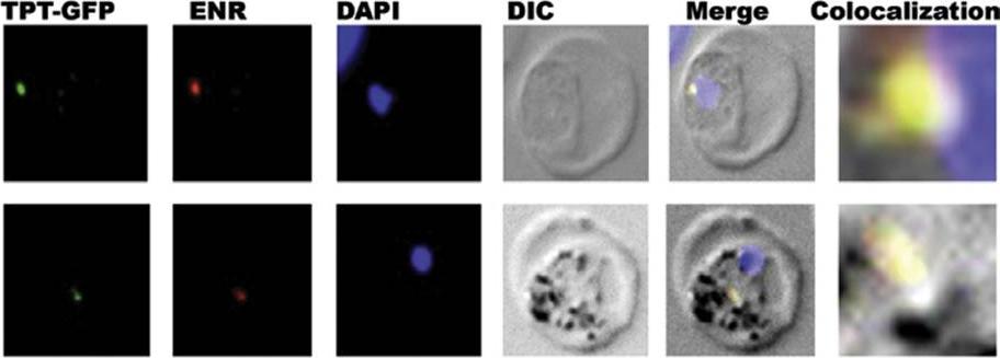
Indirect immunofluorescence assay on TPT–GFP parasites: TPT–GFP blood stage parasites were stained with primary rabbit anti-ENR polyclonal antibody and visualized by staining with secondary anti-rabbit IgG antibody coupled with Alexa 594. Parasite nuclei are stained with DAPI. In DIC are shown parasitized RBC. Merge shows overlap of TPT–GFP, rabbit anti-enoyl ACP reductase (ENR), DAPI and DIC. Apicoplast area is zoomed to show the colocalization of TPT–GFP with ENR. TPT, which was correctly localized to apicoplast. Plasmodium enoyl ACP reductase (ENR) is well-known apicoplast resident protein.Banerjee T, Jaijyan DK, Surolia N, Singh AP, Surolia A. Apicoplast triose phosphate transporter (TPT) gene knockout is lethal for Plasmodium. Mol Biochem Parasitol. 2012 186(1):44-50. . Copyright Elsevier.
See original on MMP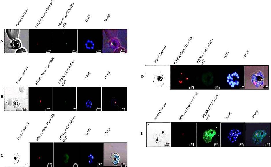
Localization ENR transit peptide double mutants to apicoplast. A field of Plasmodium cells transiently transfected with indicated ENR mutant. TufA was visualized by a rabbit anti-TufA antibody in conjugation with anti-rabbit Alexa 568 antibody (second panel) while the indicated ENR mutant was expressed in fusion with GFP (third panel). DNA was stained with DAPI (fourth panel), while the first panel displays the phase contrast image of the selected field. The fifth panel is a merge of images obtained from phase contrast, Alexa 568, GFP, and DAPI. (A) GFP-tagged ENR R40E-K42E mutant, (B) GFP-tagged ENR K42E-K49E mutant was localized to the apicoplast along with TufA, (C) GFP-tagged ENR K42A-K44A, (D) GFPtagged ENR K44A-R48A mutant, and (E) GFP-tagged ENR K51A-R52A mutant was diffusely distributed in the cytoplasmBanerjee T, Singh RR, Gupta S, Surolia A, Surolia N. 15-Deoxyspergualin hinders physical interaction between basic residues of transit peptide in PfENR and Hsp70-1. IUBMB Life. 2012 64(1):99-107.
See original on MMP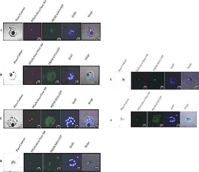
Basic residues of transit peptide essential for localization ENR to apicoplast. A field of Plasmodium cells transiently transfected with indicated PfENR mutant. TufA (encoded by the apicoplast genome) was visualized by a rabbit anti-TufA antibody in conjugation with anti-rabbit Alexa 568 antibody (second panel) while the indicated ENR mutant was expressed in fusion with GFP (third panel). DNA was stained with DAPI (fourth panel) while the first panel displays the phase constrast image of the selected field. The fifth panel is a merge of images obtained from phase contrast, Alexa 568, GFP, and DAPI. (A) GFP-tagged ENR K49A mutant was localized tothe apicoplast along with TufA, (B) GFP-tagged ENR K51A mutant was mislocalized to the cytoplasm, (C) GFP-tagged ENR R52A mutant was mislocalized to the cytoplasm, (D) GFP-tagged ENR R40A mutant was localized to the apicoplast along with TufA.(E) GFP-tagged ENR K42A mutant was localized to the apicoplast along with TufA, (F) GFP-tagged ENR K44A mutant was mislocalized to the cytoplasmBanerjee T, Singh RR, Gupta S, Surolia A, Surolia N. 15-Deoxyspergualin hinders physical interaction between basic residues of transit peptide in PfENR and Hsp70-1. IUBMB Life. 2012 64(1):99-107.
See original on MMPMore information
| PlasmoDB | PVX_113890 |
| GeneDB | PVX_113890 |
| Malaria Metabolic Pathways | Localisation images Pathways mapped to |
| Previous ID(s) | Pv113890 |
| Orthologs | PBANKA_1229800 , PCHAS_1230500 , PF3D7_0615100 , PKNH_1134900 , PVP01_1134000 , PY17X_1233300 |
| Google Scholar | Search for all mentions of this gene |