PVX_113775 6-cysteine protein (P12)
Disruptability [+]
| Species | Disruptability | Reference | Submitter | |
|---|---|---|---|---|
| P. falciparum 3D7 |
Possible |
22986493 | Theo Sanderson, Wellcome Trust Sanger Institute | |
| P. falciparum 3D7 |
Refractory |
USF piggyBac screen (Insert. mut.) | USF PiggyBac Screen | |
Mutant phenotypes [+]
None reported yet. Please press the '+' button above to add one.Imaging data (from Malaria Metabolic Pathways)
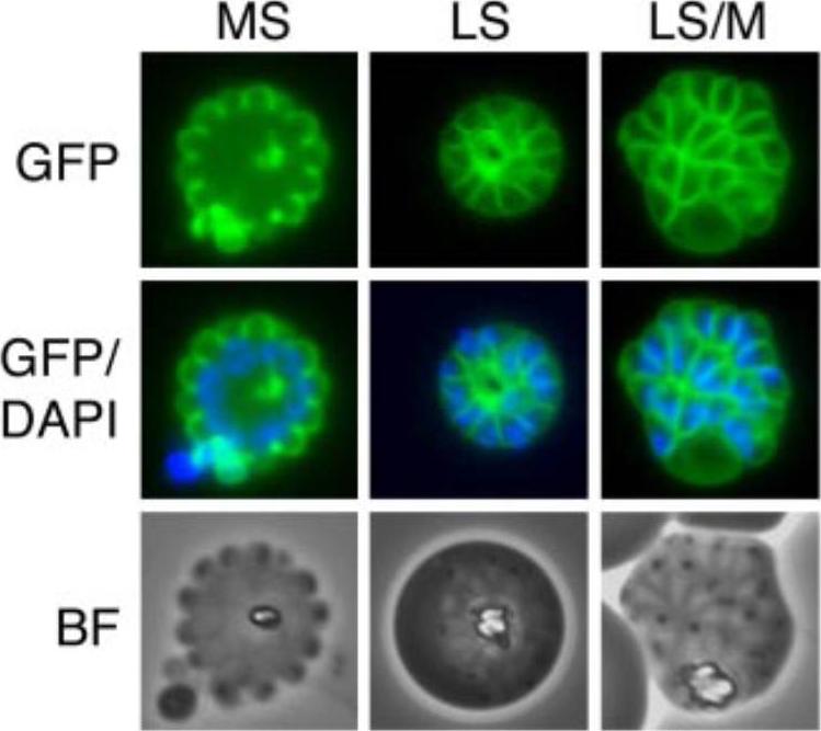
Localization of Pf12 fused to a GFP reporter and expressed in an inducible system that allows strong surface expression. MS, mid-schizont; LS, late schizont; M, merozoite. Pf12 fusion protein was localized to the merozoite surface. Pf12 is a GPI-anchored protein. Sanders PR, Gilson PR, Cantin GT, Greenbaum DC, Nebl T, Carucci DJ, McConville MJ, Schofield L, Hodder AN, Yates JR 3rd, Crabb BS. Distinct protein classes including novel merozoite surface antigens in Raft-like membranes of Plasmodium falciparum. J Biol Chem. 2005 280:40169-76.
See original on MMP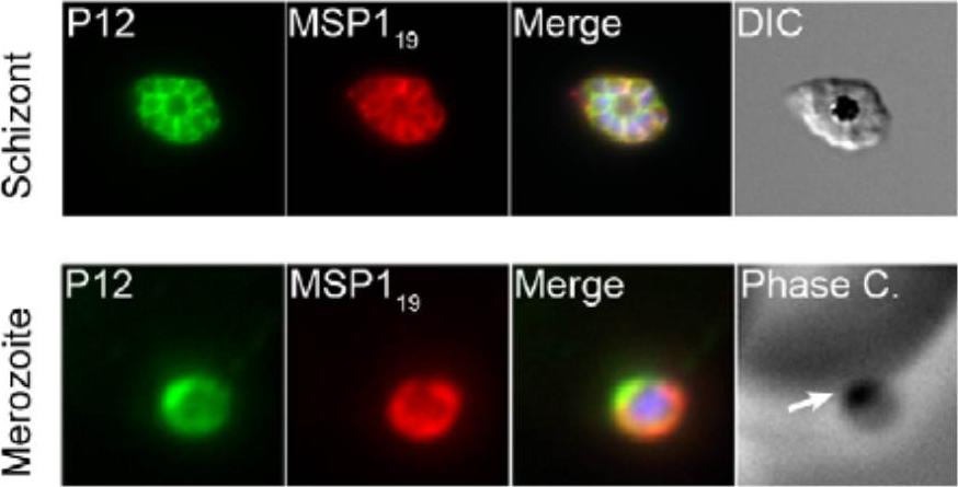
Localisation of P12 was explored by immunofluorescence assays. Schizonts and merozoites were fixed and labelled with mouse anti-MSP119 (10 mg/mL) and rabbit IgG to P12 (50 mg/mL) followed by Alexa Fluor 488 goat anti-rabbit and Alexa Fluor 568 goat anti-mouse secondary antibodies IgG. P12 was found to localise to the parasitophorous vacuole in schizonts and on the merozoite surface upon comparison with MSP119. Interestingly, P12 exhibited concentrated apical localisation on the merozoite surface. Taechalertpaisarn T, Crosnier C, Bartholdson SJ, Hodder AN, Thompson J, Bustamante LY, Wilson DW, Sanders PR, Wright GJ, Rayner JC, Cowman AF, Gilson PR, Crabb BS. Biochemical and Functional Analysis of Two Plasmodium falciparum Blood-Stage 6-Cys Proteins: P12 and P41. PLoS One. 2012;7(7):e41937.
See original on MMP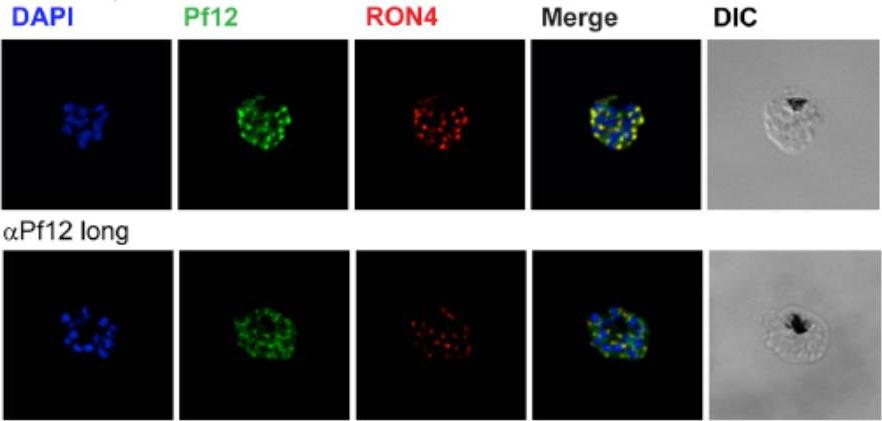
Localization of Pf12 shows punctate apical staining with anti-Pf12D2 antibody and more diffuse staining with anti-Pf12long antibody. Localization of Pf12 on mature schizonts. Confocal microscopy images of purified late-schizont infected RBCs are shown. Top panel shows labeling with affinity purified anti-Pf12D2 (against C-terminal domain), and anti-RON4 antibodies. Bottom panel shows labeling with affinity purified anti-Pf12long (against full length), and anti-RON4 antibodies. Simultaneous labeling of PfRON4, a rhoptry neck protein, consistently shows adjacent localization to Pf12 with a significant degree of co-localization. These data indicate that in the late schizont stages, Pf12 is found in an apical organelle.Tonkin ML, Arredondo SA, Loveless BC, Serpa JJ, Makepeace KA, Sundar N, Petrotchenko EV, Miller LH, Grigg ME, Boulanger MJ. Structural and biochemical characterization of Plasmodium falciparum 12 (Pf12) reveals a unique inter-domain organization and the potential for an antiparallel arrangement with Pf41. J Biol Chem. 2013 288(18):12805-17
See original on MMP
The genes for P12 and P41 can be disrupted and are therefore not essential for parasite growth. Immunofluorescence microscopy of Dp12 and Dp41 and parental 3D7 parasites probed with rabbit anti-P12 and anti-P41 IgGs in addition to the merozoite surface marker MSP1 mAb indicate absence of protein expression in the mutants aside from minor cross-reactivity.Taechalertpaisarn T, Crosnier C, Bartholdson SJ, Hodder AN, Thompson J, Bustamante LY, Wilson DW, Sanders PR, Wright GJ, Rayner JC, Cowman AF, Gilson PR, Crabb BS. Biochemical and Functional Analysis of Two Plasmodium falciparum Blood-Stage 6-Cys Proteins: P12 and P41. PLoS One. 2012;7(7):e41937.
See original on MMP
Expression and co-localization of proteins of Pfs38 complex on Plasmodium merozoites. Co-localization studies were performed by immunofluorescence assays. Merozoites were labelled with (a) mouse anti-Pfs38 and rabbit anti-GLURP or with (b) mouse anti-Pfs38 and rabbit anti-Pfs41 antibodies or with (c) mouse anti-Pfs38 and rabbit anti-Pfs12 antibodies or with (d) mouse anti-Pfs38 and rabbit anti-PfsMSP165 antibodies. Partial co-localization was observed between Pfs38 and other proteins of 6-Cys complex with co-localization coefficient of 0.75, 0.61, 0.82, and 0.83 for a–d, respectively. Td represent bright field images. Pfs38 partially co-localized with GLURP, Pfs41, Pfs12, and MSP-1, advocating the co-existence of proteins of 6-Cys protein complex on the merozoite surface (Fig. 3a–d). These results were corroborated by cosedimentation analysis of parasite-derived polypeptides. Western blotting and LC–MS/MS analysis of the glycerol gradient fractions revealed the presence of Pfs38, Pfs41, Pfs12, GLURP, MSP-1, and SERA5 in a single fraction, suggesting that these proteins exist in a complex on the parasite.Paul G, Deshmukh A, Kaur I, Rathore S, Dabral S, Panda A, Singh SK, Mohmmed A, Theisen M, Malhotra P. A novel Pfs38 protein complex on the surface of Plasmodium falciparum blood-stage merozoites. Malar J. 2017 16(1):79
See original on MMPMore information
| PlasmoDB | PVX_113775 |
| GeneDB | PVX_113775 |
| Malaria Metabolic Pathways | Localisation images Pathways mapped to |
| Previous ID(s) | Pv113775 |
| Orthologs | PBANKA_0111000 , PCHAS_0111600 , PF3D7_0612700 , PKNH_1137300 , PVP01_1136400 , PY17X_0112600 |
| Google Scholar | Search for all mentions of this gene |