PVX_084510 porphobilinogen deaminase, putative (PBGD)
Disruptability [+]
| Species | Disruptability | Reference | Submitter | |
|---|---|---|---|---|
| P. berghei ANKA |
Possible |
RMgm-1585 | Imported from RMgmDB | |
| P. berghei ANKA |
Possible |
RMgm-1577 | Imported from RMgmDB | |
| P. berghei ANKA |
Possible |
PlasmoGEM (Barseq) | PlasmoGEM | |
| P. berghei ANKA |
Possible |
RMgm-4834 | Imported from RMgmDB | |
| P. falciparum 3D7 |
Possible |
26173178 | Theo Sanderson, Wellcome Trust Sanger Institute | |
| P. falciparum 3D7 |
Refractory |
USF piggyBac screen (Insert. mut.) | USF PiggyBac Screen | |
Mutant phenotypes [+]
| Species | Stage | Phenotype | Reference | Submitter |
|---|---|---|---|---|
| P. berghei ANKA | Asexual |
No difference |
RMgm-1585 | Imported from RMgmDB |
| P. berghei ANKA | Asexual |
Difference from wild-type |
RMgm-1577
Not analysed in detail. The successful disruption of the gene encoding PBGD shows that PBGD is not essential for asexual blood stage growth/multiplication |
Imported from RMgmDB |
| P. berghei ANKA | Asexual |
No difference |
PlasmoGEM (Barseq) | PlasmoGEM |
| P. berghei ANKA | Asexual |
No difference |
RMgm-4834 | Imported from RMgmDB |
| P. berghei ANKA | Ookinete |
No difference |
RMgm-1585 | Imported from RMgmDB |
| P. berghei ANKA | Oocyst |
Difference from wild-type |
RMgm-1585
Normal numbers of oocysts are formed. However, day 10 and 14 oocysts had a reduced size (`60%) compared to wild type oocysts. A large (~10-20-fold) reduction in the numbers of midgut-associated sporozoites was observed. No hemocoel or salivary gland sporozoites were detected. |
Imported from RMgmDB |
| P. berghei ANKA | Sporozoite |
Difference from wild-type |
RMgm-1585
Normal numbers of oocysts are formed. However, day 10 and 14 oocysts had a reduced size (`60%) compared to wild type oocysts. A large (~10-20-fold) reduction in the numbers of midgut-associated sporozoites was observed. No hemocoel or salivary gland sporozoites were detected. |
Imported from RMgmDB |
| P. berghei ANKA | Liver |
Difference from wild-type |
RMgm-1585
A large (~10-20-fold) reduction in the numbers of midgut-associated sporozoites was observed. No hemocoel or salivary gland sporozoites were detected. No mosquito transmission. |
Imported from RMgmDB |
Imaging data (from Malaria Metabolic Pathways)
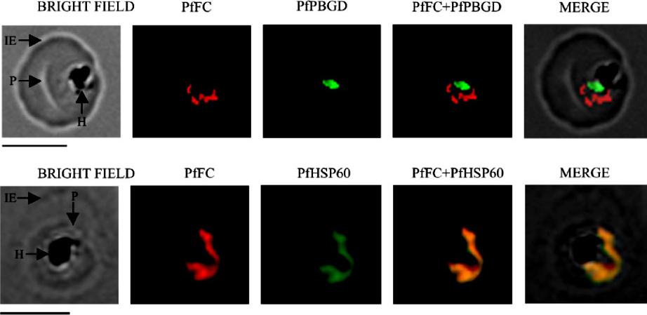
Localization of native, functional PfFC in the parasite by immunofluorescence 304 microscopy. Mouse anti-rPfFC IgG, rabbit anti-r(D)PfPBGD (apicoplast marker) IgG and rabbit anti305 rPfHSP60 (mitochondrial marker) IgG were used at 1:200 dilution. The slides were examined using a 306 Fluorescence Microscope (Leica Microsystems) and the images were acquired at 100x 307 magnification using an oil immersion objective and Leica FW 4000 image acquisition 308 software. IE, infected erythrocyte; P, parasite and H, hemozoin. Scale bar = 5 mm.Nagaraj VA, Prasad D, Rangarajan PN, Padmanaban G. Mitochondrial localization of functional ferrochelatase from Plasmodium falciparum. Mol Biochem Parasitol. 2009 168:109-12. Copyright Elsevier 2010
See original on MMP
Immunofluorescence images showing multiple localizations of PfTPxGL. PfTPxGL-red signal (anti-mouse Alexa fluor 568), PfPBGD – apicoplast marker, green signal (anti-rabbit FITC), HSP60 – mitochondrial marker, green signal (anti-rabbit FITC). The imaging was carried out with scale bars varying from 5-20 mm The infected RBCs imaged are 6-8 mm across. Parasites showing dual localization of PfTPxGL to the apicoplast and mitochondrionChaudhari R, Narayan A, Patankar S. A novel trafficking pathway in Plasmodium falciparum for the organellar localization of glutathione peroxidase-like thioredoxin peroxidase. FEBS J. 2012 279(20):3872-88
See original on MMP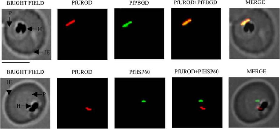
Localisation of parasite uroporphyrinogen decarboxylase (PfUROD) by immunofluorescence microscopy. Co-localisation of PfUROD with porphobilinogen deaminase (PfPBGD). Antibodies to recombinant, truncated uroporphyrinogen decarboxylase (r(D)PfUROD) were raised in mice, while PfPBGD and PfHSP60 PF10_0153 antibodies were from rabbit. To study co-localisation, tetramethylrhodamine isothiocyanate (TRITC)-conjugated goat anti-mouse IgG and FITC-conjugated goat antirabbit IgG were used. IE, infected erythrocyte; P, parasite; H, hemozoin. Scale bar = 5 mm. PfUROD co-localised with PfPBGD but not with PfHSP60 (a mitochondrial marker). Therefore, it can be concluded that PfUROD is localised in the apicoplast and not the mitochondrion.Nagaraj VA, Arumugam R, Chandra NR, Prasad D, Rangarajan PN, Padmanaban G. Localisation of Plasmodium falciparum uroporphyrinogen III decarboxylase of the heme-biosynthetic pathway in the apicoplast and characterisation of its catalytic properties. Int J Parasitol. 2009 39:559-68. Copyright Elsevier 2010.
See original on MMP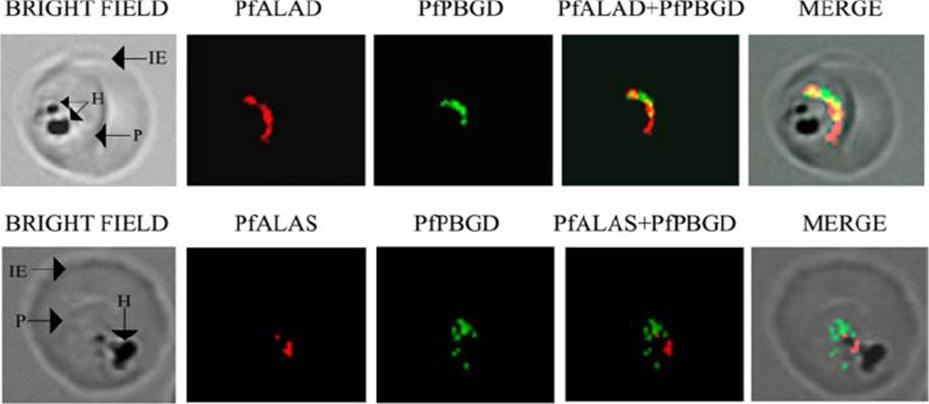
Localization of porphobilinogen deaminase PfPBGD with d-aminolevulinate dehydratase PfALAD and d-aminolevulinic acid synthase PfALAS in P. falciparum by immunofluorescence. PGDB and PfALAD are localized to the apicoplast. PfALAS is localized to the mitochondrion. IE, infected erythrocyte; P, parasite; H, hemozoin.Sato S, Clough B, Coates L, Wilson RJ. Enzymes for heme biosynthesis are found in both the mitochondrion and plastid of the malaria parasite Plasmodium falciparum. Protist. 2004 155:117-25. Copyright Elsevier 2009.
See original on MMP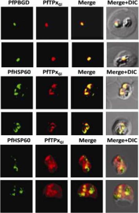
Immunofluorescence images showing multiple localizations of PfTPxGL. PfTPxGL-red signal (anti-mouse Alexa fluor 568), PfPBGD – apicoplast marker, green signal (anti-rabbit FITC), HSP60 – mitochondrial marker, green signal (anti-rabbit FITC). The imaging was carried out with scale bars varying from 5-20 mm. The infected RBCs imaged are 6-8 mm across. (Upper panel) Parasites showing only apicoplast localization of PfTPxGL, (Middle panel) Parasite showing only mitochondrial localization of PfTPxGL, (Lower panel) Parasite showing organellar as well as cytosolic localization of PfTPxGL. Chaudhari R, Narayan A, Patankar S. A novel trafficking pathway in Plasmodium falciparum for the organellar localization of glutathione peroxidase-like thioredoxin peroxidase. FEBS J. 2012 279(20):3872-88.
See original on MMP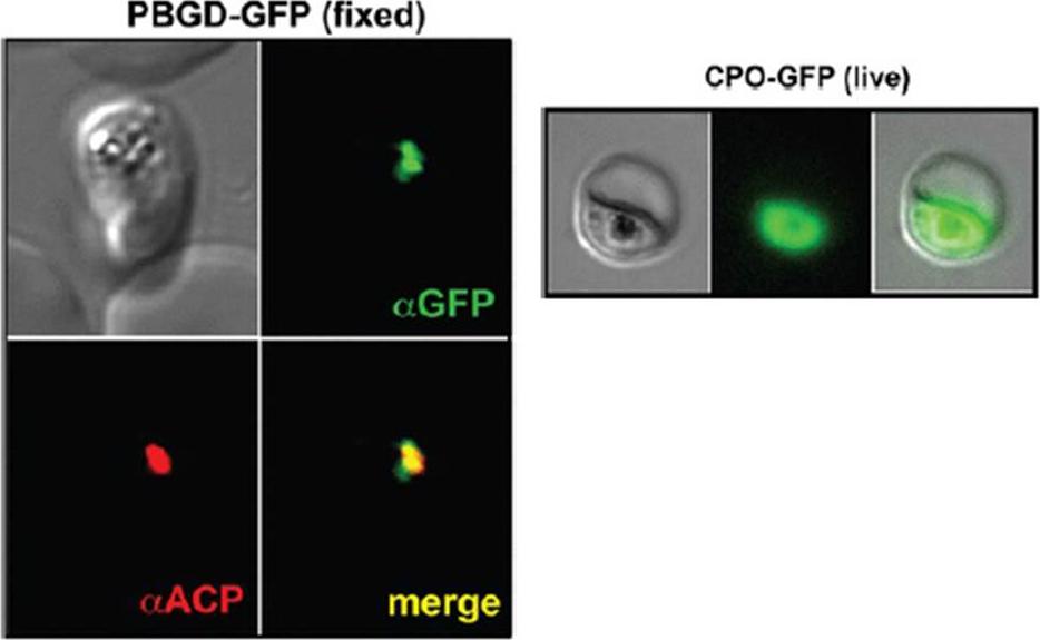
Left panel: Images of fixed 3D7 parasites expressing full-length PBGD tagged at its endogenous locus with C-terminal GFP confirm targeting of the native protein to the parasite apicoplast. Parasites were stained with αGFP and αACP (ACP, apicoplast marker).Sigala PA, Crowley JR, Henderson JP, Goldberg DE. Deconvoluting heme biosynthesis to target blood-stage malaria parasites. Elife. 2015 Jul 14;4. Right panel: Fluorescence microscopy images of live 3D7 parasites episomally expressing full-length CPO with a C-terminal GFP tag confirm protein localization to the parasite cytoplasm
See original on MMPMore information
| PlasmoDB | PVX_084510 |
| GeneDB | PVX_084510 |
| Malaria Metabolic Pathways | Localisation images Pathways mapped to |
| Previous ID(s) | Pv084510 |
| Orthologs | PBANKA_0608000 , PCHAS_0609800 , PF3D7_1209600 , PKNH_1309500 , PVP01_1308700 , PY17X_0610500 |
| Google Scholar | Search for all mentions of this gene |