PVP01_0107500 rhoptry-associated membrane antigen, putative (RAMA)
Disruptability [+]
| Species | Disruptability | Reference | Submitter | |
|---|---|---|---|---|
| P. berghei ANKA |
Refractory |
PlasmoGEM (Barseq) | PlasmoGEM | |
| P. falciparum 3D7 |
Refractory |
16790807 | Theo Sanderson, Wellcome Trust Sanger Institute | |
| P. falciparum 3D7 |
Possible |
USF piggyBac screen (Insert. mut.) | USF PiggyBac Screen | |
Mutant phenotypes [+]
| Species | Stage | Phenotype | Reference | Submitter |
|---|---|---|---|---|
| P. falciparum 3D7 | Asexual |
Invasion defect |
31491036 aberrant rhoptry morphology, secretion and invasion defects |
Abi Perrin, The Francis Crick Institute |
Imaging data (from Malaria Metabolic Pathways)
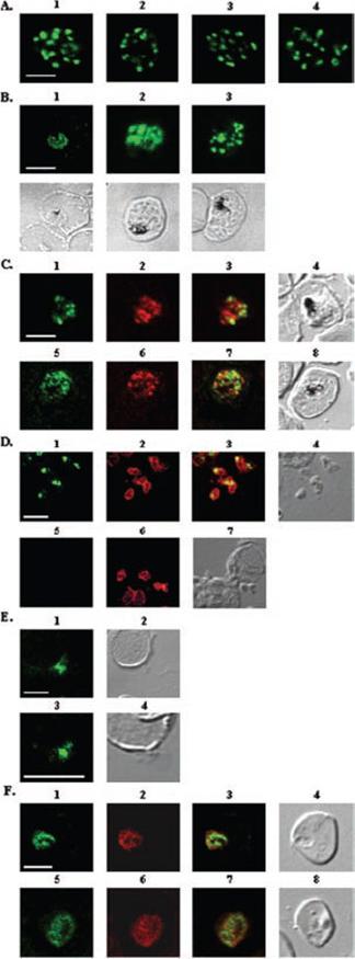
A, localization of RAMA in rhoptry organelles. Confocal microscopy on schizont stage parasites stained with rabbit anti-RAMA-B (1), anti-RAMA-C (2), anti-RAMA-D (3), or anti-RAMA-E (4) sera. B, localization of RAMA in immature parasites. Confocal microscopy on parasites stained with rabbit anti-RAMA-D serum: early trophozoite (1), late trophozoite (2), and early schizont (3) stages. Corresponding differential interference contrast (DIC) images are shown. C, trafficking of RAMA through the secretory pathway. Confocal microscopy on parasites stained with mouse anti-RAMA-E (1 and 5) and either anti-PfGRP (2) or anti-PfERD2 (6) rabbit antibodies. Corresponding overlay (3 and 7) and DIC (4 and 8) images are shown. D, localization of p60/RAMA in free merozoites. Confocal microscopy on parasites stained with mouse anti-MSP4 (2 and 6), and rabbit anti-RAMA-D (1) or anti-RAMA-B (5) sera. Corresponding overlay (3) and DIC (4 and 7) images are shown. E, discharge of RAMA from rhoptries. Confocal microscopy on CytB-treated merozoites incubated with RBCs, stained with rabbit anti-RAMA-D serum (1 and 3). Corresponding DIC images (2 and 4) are shown. F, association with the PV. Confocal microscopy on early ring stage-parasites stained with rabbit anti-RAMA-E serum (1 and 5) and monoclonal anti-RAP1 (2 and 6) antibodies. Corresponding overlay (3 and 7) and DIC (4 and 8) images are shown. Bars in 1 represent 5 mm. RAMA is synthesized as a 170-kDa protein in early trophozoites, several hours before rhoptry formation and is transiently localized within the endoplasmic reticulum and Golgi within lipid-rich microdomains.Topolska AE, Lidgett A, Truman D, Fujioka H, Coppel RL. Characterization of a membrane-associated rhoptry protein of Plasmodium falciparum. J Biol Chem. 2004 279:4648-4656.
See original on MMP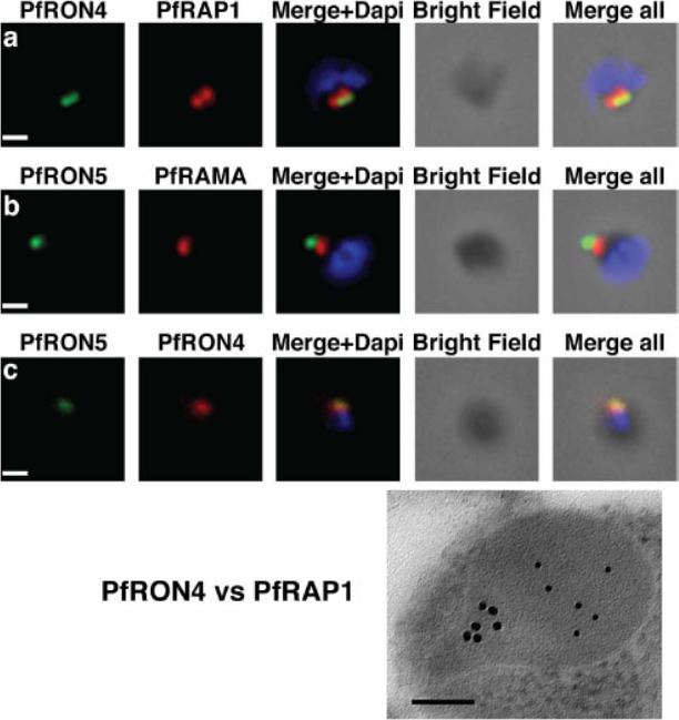
The PfRON4 and PfRON5 proteins localize to the rhoptry neck. A, Antibodies raised against PfRON4 and PfRON5 recognize specific protein products in schizont parasite extracts. B, IFAs using the anti-PfRON antibodies reveal staining of the apical tip of free merozoite, in close apposition with the rhoptry bulb markers PfRAP1 and PfRAMA, suggesting a rhoptry neck localization. Scale bar: 0.2μm C, Immunoelectron microscopy confirms that PfRON 4 localizes to the rhoptry neck. Scale bar: 0.1μmRichard D, Macraild CA, Riglar DT, Chan JA, Foley M, Baum J, Ralph SA, Norton RS, Cowman AF. Interaction between Plasmodium falciparum apical membrane antigen 1 and the Rhoptry neck protein complex defines a key step in the erythrocyte invasion process of malaria parasites. J Biol Chem. 2010 285:14815-22
See original on MMP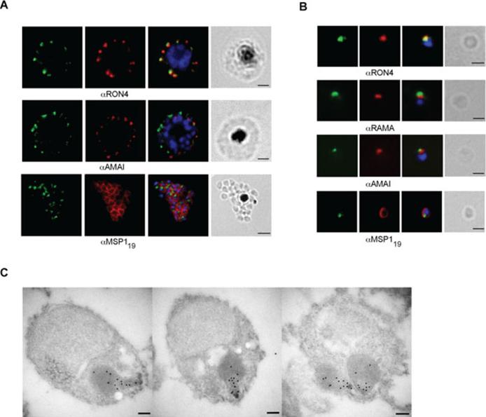
RON12 is located within the rhoptry neck.. A and B. Immunofluorescence and bright-field images of schizonts (A) and merozoites (B). Images from left to right show anti-RON12 labelling (green) followed by microneme (anti-AMA1), rhoptry (neck: anti-RON4; body: anti-RAMA) and surface (anti-MSP119) specific antibodies in red, overlay of both with DAPI-stained nuclei and the bright-field image. Size bars in (A) equal 2 μm and in (B) 1 μm. C. Immuno-electron microscopy of isolated merozoites. Localization of RON12 in the rhoptry neck of three different merozoites is shown. Size bars equal 100 nm.Knuepfer E, Suleyman O, Dluzewski AR, Straschil U, O'Keeffe AH, Ogun SA, Green JL, Grainger M, Tewari R, Holder AA. RON12, a novel Plasmodium-specific rhoptry neck protein important for parasite proliferation. Cell Microbiol. 2013 Aug 12. [Epub ahead of print]
See original on MMP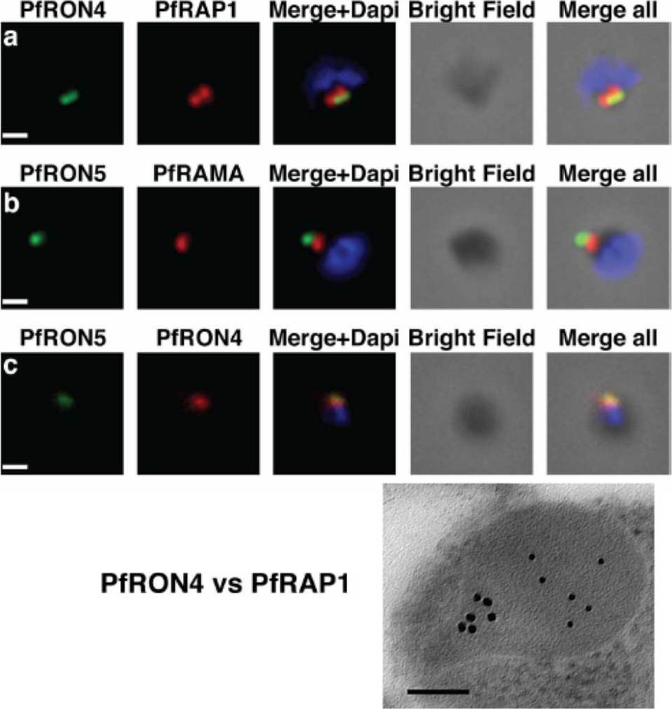
The PfRON4 and PfRON5 proteins localize to the rhoptry neck. A, Antibodies raised against PfRON4 and PfRON5 recognize specific protein products in schizont parasite extracts. B, IFAs using the anti-PfRON antibodies reveal staining of the apical tip of free merozoite, in close apposition with the rhoptry bulb markers PfRAP1 and PfRAMA, suggesting a rhoptry neck localization. Scale bar: 0.2μm C, Immunoelectron microscopy confirms that PfRON 4 localizes to the rhoptry neck. Scale bar: 0.1μmRichard D, Macraild CA, Riglar DT, Chan JA, Foley M, Baum J, Ralph SA, Norton RS, Cowman AF. Interaction between Plasmodium falciparum apical membrane antigen 1 and the Rhoptry neck protein complex defines a key step in the erythrocyte invasion process of malaria parasites. J Biol Chem. 2010 285:14815-22
See original on MMP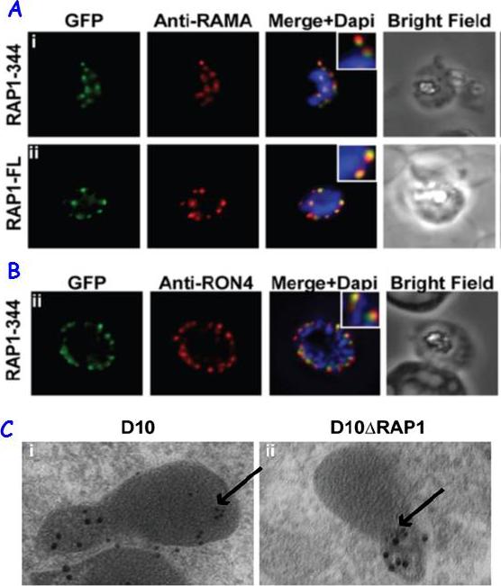
RAP1 contains a bipartite rhoptry signal. A RAP1-344 and RAP1-FL GFP fusions. Both constructs show a punctate fluorescence pattern characteristic of rhoptry localisation. For the RAP1-FL construct, GFP signal overlaps with RAMA. In contrast, the RAP1-344GFP chimera only partially overlaps with RAMA, suggesting rhoptry neck localisation. B. For the RAP1-344 construct, GFP co-localises with the rhoptry neck marker PfRON4. C. Immunoelectron microscopy demonstrates that truncated RAP1 (10 nm beads) in D10DRAP1 parasites is localised in the rhoptry neck, whereas full-length RAP1 in D10 (wild-type) parasites is localised in the rhoptry bulb. PfRON4 PF11_0168 (15 nm beads) is localised in the rhoptry neck in both parasite lines.Richard D, Kats LM, Langer C, Black CG, Mitri K, Boddey JA, Cowman AF, Coppel RL. Identification of Rhoptry Trafficking Determinants and Evidence for a Novel Sorting Mechanism in the Malaria Parasite Plasmodium falciparum. PLoS Pathog. 2009 5(3):e1000328.
See original on MMP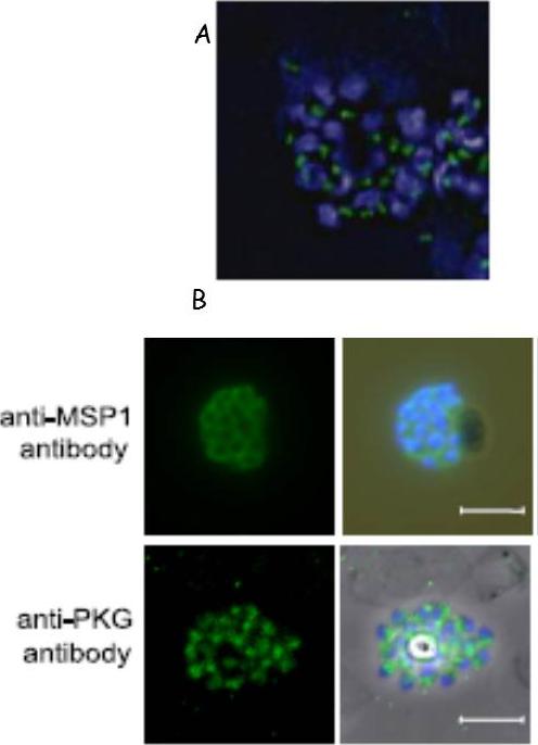
Fluorescence micrographs of schizonts. The primary antibody was a monoclonal antibody that reacts with the P. falciparum rhoptry neck protein PfRON4 (green). Nuclei were counterstained with 0.5 g/ml DAPI (blue). Scale bar, 5 mm. (B) Fluorescence micrographs of schizonts. The primary antibodies were a monoclonal antibody that reacts with P. falciparum MSP1 (top row) and a polyclonal antibody that reacts with PfPKG (bottom row) (both green). Nuclei were counterstained with 0.5 g/ml DAPI (blue). The staining pattern observed throughout the merozoites suggests a predominantly cytosolic localization.Taylor HM, McRobert L, Grainger M, Sicard A, Dluzewski AR, Hopp CS, Holder AA, Baker DA. The malaria parasite cyclic GMP-dependent protein kinase plays a central role in blood-stage schizogony. Eukaryot Cell. 2010 9:37-45.
See original on MMP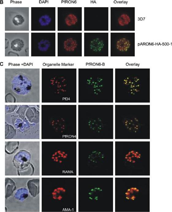
Localisation of the PfRON6 protein in parasitised red blood cells. (B) Double labelling using anti-rPfRON6-B and anti- haemagglutinin (HA) in HA-tagged transgenic parasites. Confocal microscopy using anti-rPfRON6-B with anti-HA. Corresponding overlay images are shown. (C) Double labelling of PfRON6 with Pf34, PfRON4, RAMA or AMA1 in segmented schizonts. Confocal microscopy using anti-rPfRON6-B with anti-Pf34 (rhoptry neck marker), anti-PfRON4 (rhoptry neck marker), anti-RAMA (rhoptry bulb marker), or anti-AMA1 (microneme marker). Corresponding overlay images are shown. a punctate pattern of fluorescence in segmented schizonts characteristic of localisation with the apical secretory organelles.Proellocks NI, Kats LM, Sheffield DA, Hanssen E, Black CG, Waller KL, Coppel RL. Characterisation of PfRON6, a Plasmodium falciparum rhoptry neck protein with a novel cysteine-rich domain. Int J Parasitol. 2008 39(6):683-92. Copyright Elsevier 2009.
See original on MMP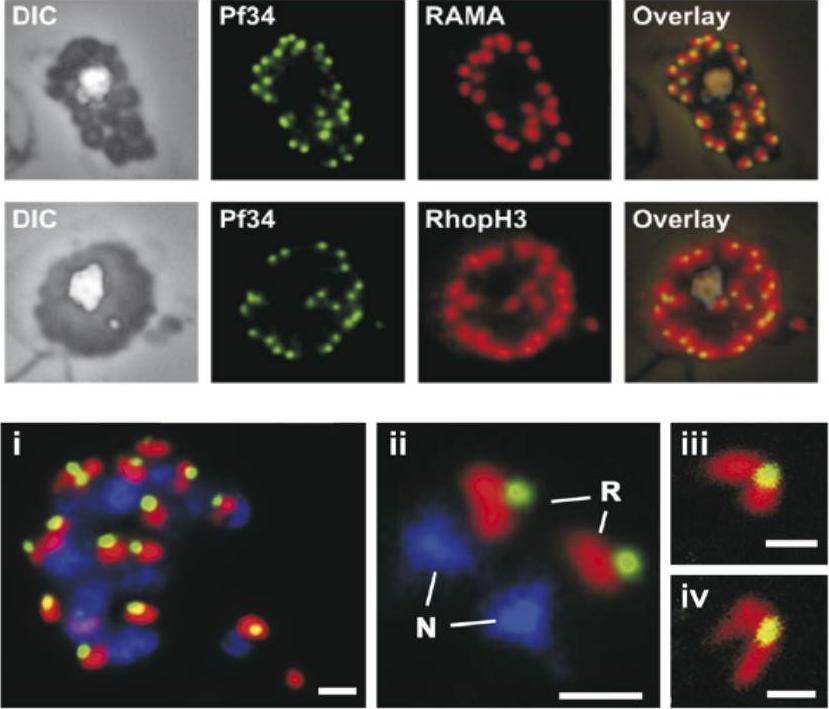
Upper 2 rows: Localization of Pf34 to the rhoptries was demonstrated in late stage parasites by immunostaining infected blood smears with anti-Pf34 and co-localising with either anti-RAMA or anti-RhopH3 (both rhoptry markers) or anti-AMA-1 (microneme marker) antibodies.Lower row: A mature schizont (i) and two extracellular merozoites (ii) showing the close apposition of Pf34-positive (green) and RAMA-positive (red) structures in the apex of the merozoite. Panels (iii) and (iv) show the apex of merozoites and the two rhoptries connected to the Pf34-positive structure at the anterior when visualised by confocal microscopy. N, nucleus; R, rhoptry. Bars represent 1 mm in (i) and (ii) and 500 nm in (iii) and (iv).Proellocks NI, Kovacevic S, Ferguson DJ, Kats LM, Morahan BJ, Black CG, Waller KL, Coppel RL. Plasmodium falciparum Pf34, a novel GPI-anchored rhoptry protein found in detergent-resistant microdomains. Int J Parasitol. 2007 37:1233-41. Copyright Elsevier
See original on MMP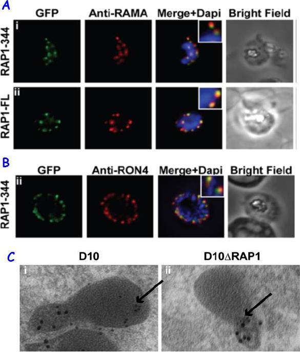
RAP1 contains a bipartite rhoptry signal. A RAP1-344 and RAP1-FL GFP fusions. Both constructs show a punctate fluorescence pattern characteristic of rhoptry localisation. For the RAP1-FL construct, GFP signal overlaps with RAMA. In contrast, the RAP1-344GFP chimera only partially overlaps with RAMA, suggesting rhoptry neck localisation. B. For the RAP1-344 construct, GFP co-localises with the rhoptry neck marker PfRON4. C. Immunoelectron microscopy demonstrates that truncated RAP1 (10 nm beads) in D10DRAP1 parasites is localised in the rhoptry neck, whereas full-length RAP1 in D10 (wild-type) parasites is localised in the rhoptry bulb. PfRON4 PF11_0168 (15 nm beads) is localised in the rhoptry neck in both parasite lines.Richard D, Kats LM, Langer C, Black CG, Mitri K, Boddey JA, Cowman AF, Coppel RL. Identification of Rhoptry Trafficking Determinants and Evidence for a Novel Sorting Mechanism in the Malaria Parasite Plasmodium falciparum. PLoS Pathog. 2009 5(3):e1000328.
See original on MMP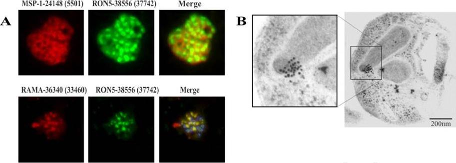
PfRON5 localizes to the rhoptry neck. (A) Aotus antibodies against MSP-1 peptide 24148 (5501) displaying the membrane, and against RAMA peptide 36340 (33460),showing the rhoptries (red fluorescence), together with RON5 peptide 38556 (37742) (green fluorescence) and DAPI (blue fluorescence, merged at the bottom), confirming RON5 localization in the rhoptries but differently to that of RAMA. (B) Immuno-electron microscopy with rabbit antibodies against RON5 peptides and protein A-gold showed specific labeling in the P. falciparum merozoite rhoptry neck. PfRON5 was detected in pear-shaped rhoptry neck portion in segmented schizonts when using rabbit polyclonal antibodies for IEM assay.Curtidor H, Patiño LC, Arévalo-Pinzón G, Vanegas M, Patarroyo ME, Patarroyo MA. Plasmodium falciparum rhoptry neck protein 5 peptides bind to human red blood cells and inhibit parasite invasion. Peptides. 2013 Aug 8. 9781(13)00268-4.
See original on MMP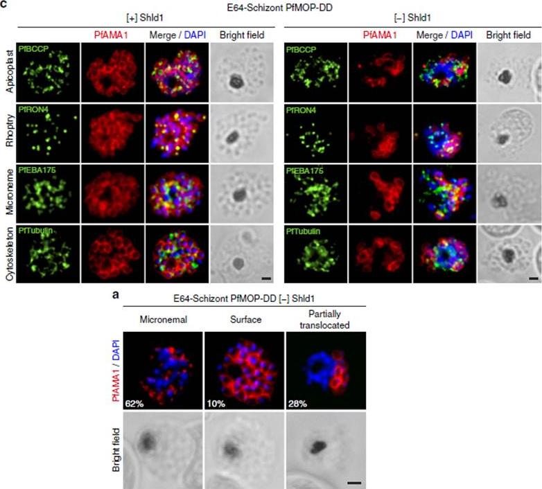
PfAMA1 translocation is aberrant in schizonts with PfMOP-knockdown. (a) Schizonts from [+]/[-] Shld1 PfMOP-DD parasites were E64-treated, fixed, probed with anti-PfAMA1 and scored as micronemal (M), partially translocated (PT) or surface (S), representative [+] Shld1 parasite IFAs shown. (c) Synchronized schizont stage (40–44 h) parasites, maintained with 250nM (left panel) or 0 nM (right panel) Shld1, were incubated 6 h in presence of 10 mM E64, methanol-fixed, permeabilized, and stained using antibodies against PfRON4, PfRhopH3, PfEBA175 and PfTubulin. Staining for these markers was similar in [+] and [-] Shld1 conditions. PfAMA1 staining was used to identify E64-treated schizonts that were sufficiently mature (that is, surface staining or partially translocated staining, but not micronemal staining, scale bar, 1 mm).Absalon S, Robbins JA, Dvorin JD. An essential malaria protein defines the architecture of blood-stage and transmission-stage parasites. Nat Commun. 2016 7:11449.
See original on MMP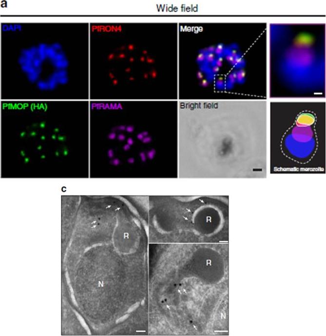
PfMOP localizes to apical area of parasites. Representative pictures of E64-treated schizont-stages. (a) Wide-field IFA of segmented schizont stained with antibodies against PfRAMA, PfRON4, and HA. Scale bar, 1 mm (c) Transmission electron microscopy with (white arrows) gold-labelled antibodies against HA (N, nucleus; R, rhoptry, scale bar, 100 nm).Absalon S, Robbins JA, Dvorin JD. An essential malaria protein defines the architecture of blood-stage and transmission-stage parasites. Nat Commun. 2016 7:11449.
See original on MMP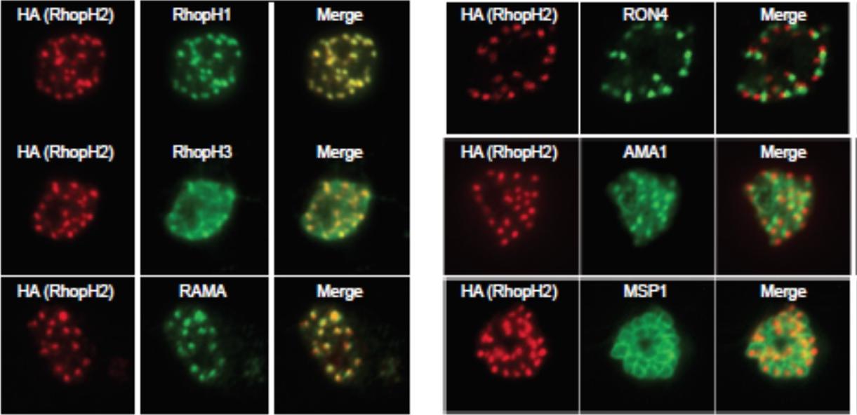
Generation of transgenic parasites in which RhopH2 is epitope-tagged. Immunofluorescence analysis (IFA) on schizonts fixed with acetone/methanol and labelled with anti-HA antibody to detect RhopH2 and other antibodies, as indicated. Immunofluorescence analysis (IFA) confirmed RhopH2-HA localized to the rhoptry and co-localized with other rhoptry bulb proteins, RhopH1, RhopH3 and RAMA but not with the rhoptry neck protein, RON4, the micronemal marker, AMA-1 or the plasma membrane protein MSP1.Counihan N, Chisholm SA, Bullen HE, Srivastava A, Sanders PR, Jonsdottir TK, Weiss GE, Ghosh S, Crabb BS, Creek DJ, Gilson PR, de Koning-Ward TF. Plasmodium falciparum parasites deploy RhopH2 into the host erythrocyte to obtain nutrients, grow and replicate. Elife. 2017 Mar 2;6. pii: e23217
See original on MMPMore information
| PlasmoDB | PVP01_0107500 |
| GeneDB | PVP01_0107500 |
| Malaria Metabolic Pathways | Localisation images Pathways mapped to |
| Previous ID(s) | null |
| Orthologs | PBANKA_0804500 , PCHAS_0804800 , PF3D7_0707300 , PKNH_0105800 , PVX_087885 , PY17X_0807600 |
| Google Scholar | Search for all mentions of this gene |