PKNH_1106400 ferrochelatase, putative (FC)
Disruptability [+]
| Species | Disruptability | Reference | Submitter | |
|---|---|---|---|---|
| P. berghei ANKA |
Possible |
RMgm-1578 | Imported from RMgmDB | |
| P. berghei ANKA |
Possible |
RMgm-925 | Imported from RMgmDB | |
| P. berghei ANKA |
Possible |
RMgm-4175 | Imported from RMgmDB | |
| P. berghei ANKA |
Possible |
RMgm-5263 | Imported from RMgmDB | |
| P. falciparum 3D7 |
Possible |
25352601 | Theo Sanderson, Wellcome Trust Sanger Institute | |
| P. falciparum 3D7 |
Possible |
USF piggyBac screen (Insert. mut.) | USF PiggyBac Screen | |
Mutant phenotypes [+]
| Species | Stage | Phenotype | Reference | Submitter |
|---|---|---|---|---|
| P. berghei ANKA | Asexual |
No difference |
RMgm-925 | Imported from RMgmDB |
| P. berghei ANKA | Asexual |
No difference |
RMgm-4175 | Imported from RMgmDB |
| P. berghei ANKA | Asexual |
Difference from wild-type |
RMgm-5263
ECM-susceptible C57BL/6 were by injected with 10(5) asexual stage parasites. Blood parasitemia showed 23 days delay in the growth of FC-KO parasites with respect to the wild type (WT). About 80% of the WT-infected mice succumbed to ECM within day 10 when the blood parasitemia was around 1030%. In contrast, mice infected with ALASKO and FCKO parasites were protected from ECM and they died because of anemia on day 2030. |
Imported from RMgmDB |
| P. falciparum 3D7 | Asexual |
No difference |
25352601 | Theo Sanderson, Wellcome Trust Sanger Institute |
| P. berghei ANKA | Gametocyte |
No difference |
RMgm-925 | Imported from RMgmDB |
| P. berghei ANKA | Gametocyte |
No difference |
RMgm-4175 | Imported from RMgmDB |
| P. falciparum 3D7 | Gametocyte |
Attenuated |
25352601 | Theo Sanderson, Wellcome Trust Sanger Institute |
| P. berghei ANKA | Ookinete |
No difference |
RMgm-925 | Imported from RMgmDB |
| P. berghei ANKA | Ookinete |
No difference |
RMgm-4175 | Imported from RMgmDB |
| P. berghei ANKA | Oocyst |
Difference from wild-type |
RMgm-925
Strongly reduced oocyst production |
Imported from RMgmDB |
| P. berghei ANKA | Oocyst |
Difference from wild-type |
RMgm-4175
Oocysts were produced but no salivary gland sporozoites |
Imported from RMgmDB |
| P. falciparum 3D7 | Oocyst |
Attenuated |
25352601 | Theo Sanderson, Wellcome Trust Sanger Institute |
| P. berghei ANKA | Sporozoite |
Difference from wild-type |
RMgm-925
Strongly reduced oocyst and no sporozoite production.Mice remained negative when infected by mosquito bite or after i.v. injection of sporozoites (see also additional remarks phenotype) |
Imported from RMgmDB |
| P. berghei ANKA | Sporozoite |
Difference from wild-type |
RMgm-4175
Oocysts were produced but no salivary gland sporozoites |
Imported from RMgmDB |
| P. berghei ANKA | Liver |
Difference from wild-type |
RMgm-925
Strongly reduced oocyst and no sporozoite production. |
Imported from RMgmDB |
| P. berghei ANKA | Liver |
Difference from wild-type |
RMgm-4175
See additional information on the generation and analysis of sporozoites lacking the FC gene (these FC(KO) sporozoites are motile and liver infective both in vitro and in vivo but have severe growth defects and fail to complete liver stage development) |
Imported from RMgmDB |
Imaging data (from Malaria Metabolic Pathways)

1. Analysis of the co-localization of PfFC with PfHsp60 using FITC-conjugated (green) secondary antibodies for the former and TRITC-conjugated (red) secondary antibodies for the latter; 1, bright field (dark spots represent the hemozoin pigment); 2, red fluorescence in the parasite due to PfHsp60; 3, green fluorescence in the parasite due to PfFC; 4, merge of 2 and 3 along with Hoescht stain (blue) showing lack of co-localization; 5, projection of two parasites showing lack of co-localization.2. Three different fields showing discrete gold particles in typical apicoplast structures with multiple membranes. 2 part of the whole parasite; 3, elongating apicoplasts in a later-stage trophozoite; 4, a circular apicoplast in an early-stage tropozoite. The signals are localized close to the membrane. Scale bar, 100 μm. AP, apicoplast; M, mitochondrion; R, red cell; MC, Maurer’s cleft; P, parasite; Pv, parasite food vacuole; Pc, parasite cytosol.Reproduced with permission, from Varadharajan S, Sagar BK, Rangarajan PN, Padmanaban G. Localization of ferrochelatase in Plasmodium falciparum. Biochem J. 2004 384:429-36. © the Biochemical Society
See original on MMP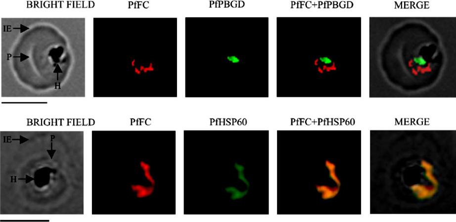
Localization of native, functional PfFC in the parasite by immunofluorescence 304 microscopy. Mouse anti-rPfFC IgG, rabbit anti-r(D)PfPBGD (apicoplast marker) IgG and rabbit anti305 rPfHSP60 (mitochondrial marker) IgG were used at 1:200 dilution. The slides were examined using a 306 Fluorescence Microscope (Leica Microsystems) and the images were acquired at 100x 307 magnification using an oil immersion objective and Leica FW 4000 image acquisition 308 software. IE, infected erythrocyte; P, parasite and H, hemozoin. Scale bar = 5 mm.Nagaraj VA, Prasad D, Rangarajan PN, Padmanaban G. Mitochondrial localization of functional ferrochelatase from Plasmodium falciparum. Mol Biochem Parasitol. 2009 168:109-12. Copyright Elsevier 2010
See original on MMP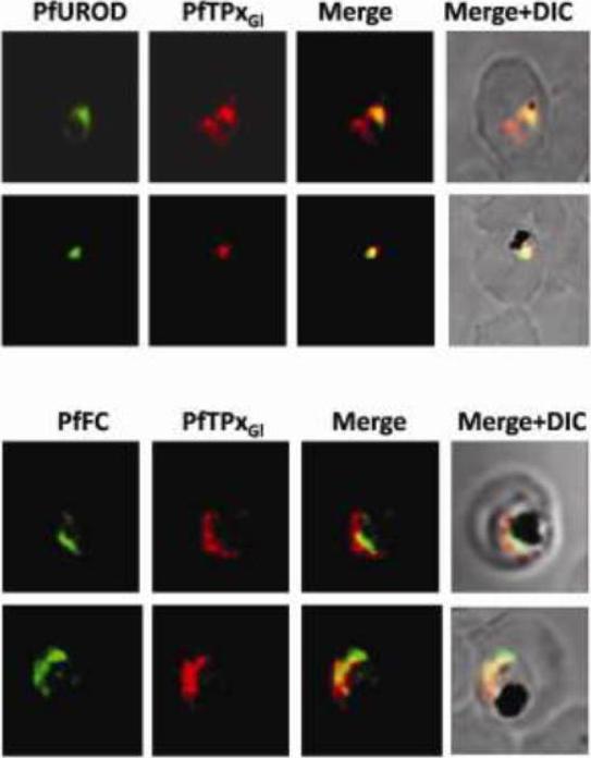
Immunofluorescence images showing multiple localizations of PfTPxGL. PfTPxGL-red signal (anti-mouse Alexa fluor 568). The imaging was carried out with scale bars varying from 5-20 mm. The infected RBCs imaged are 6-8 mm across. Parasites showing localization of PfTPxGl using antibodies purified against rPfTPxGL. In several parasites, PfTPxGL was localized exclusively to only one organelle per parasite. In some parasites, PfTPxGL co-localized with exclusively the mitochondrial markers PfUROD or PfFC. Chaudhari R, Narayan A, Patankar S. A novel trafficking pathway in Plasmodium falciparum for the organellar localization of glutathione peroxidase-like thioredoxin peroxidase. FEBS J. 2012 279(20):3872-88 PMID:
See original on MMP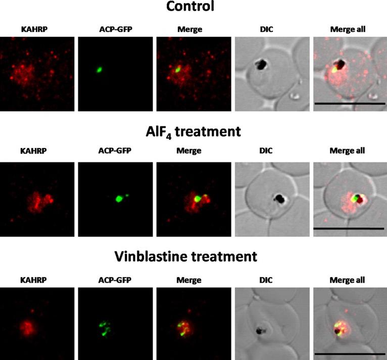
Immunofluorescence images showing PfTPxGl and PfFC (mitochondrial marker protein) trafficking in AlF4−, nocodazole and vineblsatine treated 3D7 parasites(A) PfTPxGl and PfFC co-localization in AlF4−-treated parasites, (B) PfTPxGl and PfFC co-localization in vinblastine-treated parasites, (C) PfTPxGl and PfFC co-localization in nocodazole-treated parasites. In this experiment, PfTPxGl was found to be co-localized with the mitochondrial marker protein PfFC in 40% of the treated parasites suggesting that trafficking of PfTPxGl to the mitochondrion may be partially disrupted by the treatments. Scale Bar: 10 µm. When the parasites from the same treated cultures were analyzed for co-localization of the disrupted PfTPxGl signal with mitochondrial marker protein ferrochelatase (PfFC), we observed that in 35-40% of cells the PfTPxGl signal showed some overlap.Chaudhari R, Dey V, Narayan A, Sharma S, Patankar S. Membrane and luminal proteins reach the apicoplast by different trafficking pathways in the malaria parasite Plasmodium falciparum. PeerJ. 2017 Apr 27;5:e3128. PMID: 28462015
See original on MMPMore information
| PlasmoDB | PKNH_1106400 |
| GeneDB | PKNH_1106400 |
| Malaria Metabolic Pathways | Localisation images Pathways mapped to |
| Previous ID(s) | PK13_6630c, PKH_110580 |
| Orthologs | PBANKA_1140700 , PCHAS_1140200 , PF3D7_1364900 , PVP01_1107100 , PVX_115205 , PY17X_1142100 |
| Google Scholar | Search for all mentions of this gene |