PF3D7_1479000 acyl-CoA synthetase (ACS1a)
Disruptability [+]
| Species | Disruptability | Reference | Submitter |
|---|---|---|---|
| P. falciparum 3D7 |
Refractory |
USF piggyBac screen (Insert. mut.) | USF PiggyBac Screen |
Mutant phenotypes [+]
None reported yet. Please press the '+' button above to add one.Imaging data (from Malaria Metabolic Pathways)
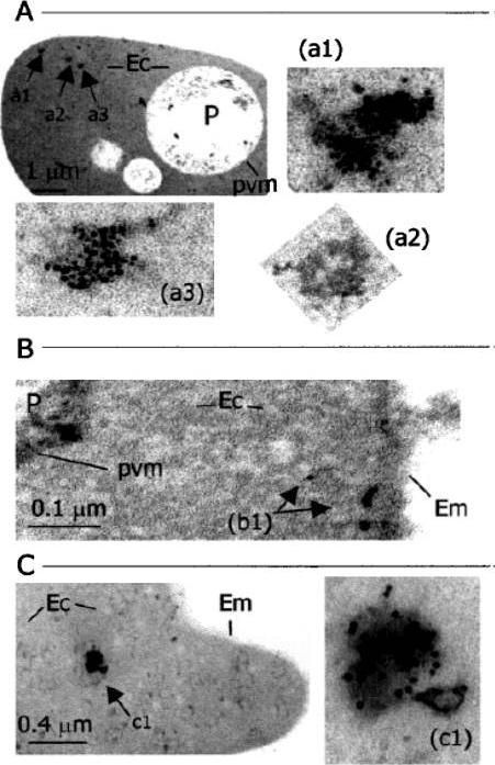
Immunoelectron microscopy of P. falciparum-infected erythrocytes labeled with anti-15 antibody and protein G-gold (particles of 10 nm diameter). Arrows in the photomicrogaphs indicate different structures containing PfACS1 protein located in the erythrocyte cytoplasm (Ec) and outside the intracellular parasite (P) in all the cases observed. In structures (a1) and (a3), the PfACS1 appears to be located inside the vesicle or as possible protein aggregates; (a2) is a vesicle-like structure that did not show labeling with the anti-15 antibody; (c1) shows apparent labeling on the surface of membrane structures of this vesicle; and (b1) is a linear string of labeling close of the erythrocyte membrane (Em). Pvm signifies parasitophorous vacuole membrane. All the photographs were taken with a Zeiss electronmicroscope at (a) 10,000x and (b) and (c) 50,000x. The negative films were scanned-digitalized and the images processed with Adobe Photoshop to get the best contrast.Matesanz F, Durán-Chica I, Alcina A. The cloning and expression of Pfacs1, a Plasmodium falciparum fatty acyl coenzyme A synthetase-1 targeted to the host erythrocyte cytoplasm. J Mol Biol. 1999 291:59-70.
See original on MMPMore information
| PlasmoDB | PF3D7_1479000 |
| GeneDB | PF3D7_1479000 |
| Malaria Metabolic Pathways | Localisation images Pathways mapped to |
| Previous ID(s) | PF14_0761 |
| Orthologs | |
| Google Scholar | Search for all mentions of this gene |