PF3D7_1473700 nucleoporin NUP116/NSP116, putative (NUP116)
Disruptability [+]
| Species | Disruptability | Reference | Submitter |
|---|---|---|---|
| P. falciparum 3D7 |
Possible |
USF piggyBac screen (Insert. mut.) | USF PiggyBac Screen |
Mutant phenotypes [+]
None reported yet. Please press the '+' button above to add one.Imaging data (from Malaria Metabolic Pathways)
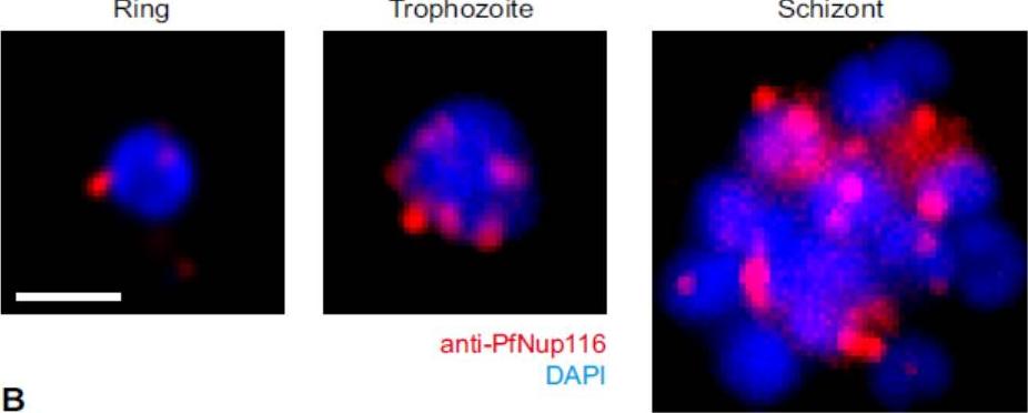
Nuclear pore redistribution throughout the intraerythrocytic stages. Immuno-fluorescence staining of nuclear pores using anti-PfNup116 at different intra-erythrocytic stages. Schizont stage image is a maximum ntensity projection. Nucleus stained by DAPI. Bar, 1 μm. Guizetti J, Martins RM, Guadagnini S, Claes A, Scherf A. Nuclear pores and perinuclear expression sites of var and rDNA genes correspond to physically distinct regions in Plasmodium falciparum. Eukaryot Cell. 2013 12(5):697-702.
See original on MMP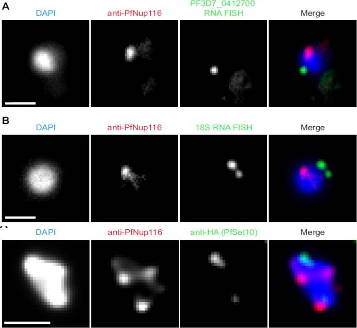
Upper 2 panels: Colocalization of gene expression sites with nuclear pores. (A) Combination of anti-PfNup116 immuno-fluorescence staining with RNA-FISH using a probe against the active var gene expressed in 3D7-G7 clone (PF3D7_0412700). Nucleus stained by DAPI. Bar, 1 μm.(B) As in (A) with FISH probe against 18S ribosomal RNA. (C) Fraction of cells with an overlap of FISH and nuclear pore foci. Lower panel: Colocalization of PfSet10-HA with nuclear pores. Combination of anti-PfNup116 and anti-HA immunofluorescence staining on early trophozoite stage parasite expressing recombinant PfSet10-HA. Nucleus stained by DAPI. Bar, 1 μm.Guizetti J, Martins RM, Guadagnini S, Claes A, Scherf A. Nuclear pores and perinuclear expression sites of var and rDNA genes correspond to physically distinct regions in Plasmodium falciparum. Eukaryot Cell. 2013 12(5):697-702
See original on MMP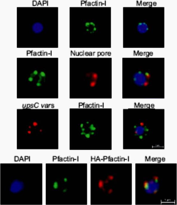
Upper panel: IFA visualization of subcellular actin in rings using an anti-Pfactin-I antibody (rabbit #2). Second panel: Two-color IFA assay using anti-Pfactin-I (rabbit #2, green) and anti-nuclear pore (PF14_0706) antibody (rat, red). Third panel: Combined FISH/IFA assay in rings showing partial colocalization of chromosome internal var genes (upsC-type) and actin. Fourth panel: IFA analysis of HA-actin-transfected ring stage parasites using mouse anti-HA antibody (red) and anti-Pfactin-I antibody (rabbit #2, green). IFA analysis with anti-Pfactin-I antibodies or with anti-HA antibody in HAactin-transfected 3D7 parasites confirmed the restricted location of actin to the nucleus in ring stage parasites and demonstrated that it concentrated in foci at the nuclear periphery. Colocalization with a nuclear pore antibody indicates that the staining is limited to the inner side of the nucleus.Zhang Q, Huang Y, Zhang Y, Fang X, Claes A, Duchateau M, Namane A, Lopez-Rubio JJ, Pan W, Scherf A. A critical role of perinuclear ilamentous actin in spatial repositioning and mutually exclusive expression of virulence genes in malaria parasites. Cell Host Microbe. 2011 10:451-63.
See original on MMPMore information
| PlasmoDB | PF3D7_1473700 |
| GeneDB | PF3D7_1473700 |
| Malaria Metabolic Pathways | Localisation images Pathways mapped to |
| Previous ID(s) | PF14_0706 |
| Orthologs | |
| Google Scholar | Search for all mentions of this gene |