PF3D7_1462800 glyceraldehyde-3-phosphate dehydrogenase (GAPDH)
Disruptability [+]
| Species | Disruptability | Reference | Submitter | |
|---|---|---|---|---|
| P. falciparum 3D7 |
Refractory |
USF piggyBac screen (Insert. mut.) | USF PiggyBac Screen | |
| P. berghei ANKA |
Refractory |
PlasmoGEM (Barseq) | PlasmoGEM | |
Mutant phenotypes [+]
None reported yet. Please press the '+' button above to add one.Imaging data (from Malaria Metabolic Pathways)
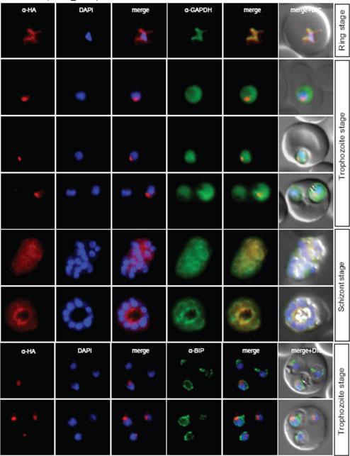
Localisation of NuProC2-3xHA (PF10_0278) during the IDC. Localisation of the tagged protein was visualised using anti-HA antibodies (red). Antibodies against GAPDH and PfBIP were used to visualise the cytosolic (top panel) and ER (bottom panel) compartments, respectively. DAPI was used to visualise the nucleus. DIC images are shown as reference. The protein co-localized with fibrillarin to the parasite nucleolus.Oehring SC, Woodcroft BJ, Moes S, Wetzel J, Dietz O, Pulfer A, Dekiwadia C, Maeser P, Flueck C, Witmer K, Brancucci NM, Niederwieser I, Jenoe P, Ralph SA, Voss TS. Organellar proteomics reveals hundreds of novel nuclear proteins in the malaria parasite Plasmodium falciparum. Genome Biol. 2012 Nov 26;13(11):R108.
See original on MMP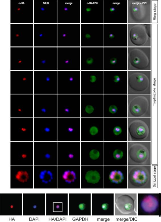
Localisation of PF10_0328 during the IDC. Localisation of the tagged protein was visualised using anti-HA antibodies (red). Antibodies against GAPDH were used to visualise the cytosolic compartment. DAPI was used to visualise the nucleus. DIC images are shown as reference. The protein localized to unknown nuclear subcompartment.Oehring SC, Woodcroft BJ, Moes S, Wetzel J, Dietz O, Pulfer A, Dekiwadia C, Maeser P, Flueck C, Witmer K, Brancucci NM, Niederwieser I, Jenoe P, Ralph SA, Voss TS. Organellar proteomics reveals hundreds of novel nuclear proteins in the malaria parasite Plasmodium falciparum. Genome Biol. 2012 Nov 26;13(11):R108.
See original on MMP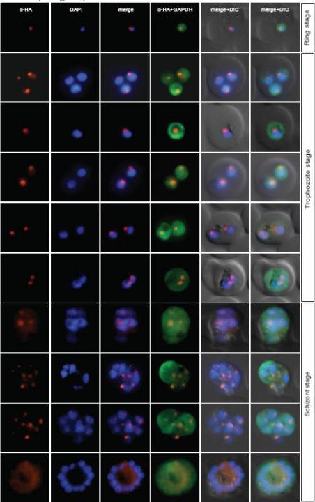
Localisation of NuProC3-3xHA (PF11_0250) during the IDC. Localisation of the tagged protein was visualised using anti-HA antibodies (red). Antibodies against GAPDH were used to visualise the cytosolic compartment. DAPI was used to visualise the nucleus. DIC images are shown as reference. The protein co-localized with fibrillarin to the parasite nucleolus.Oehring SC, Woodcroft BJ, Moes S, Wetzel J, Dietz O, Pulfer A, Dekiwadia C, Maeser P, Flueck C, Witmer K, Brancucci NM, Niederwieser I, Jenoe P, Ralph SA, Voss TS. Organellar proteomics reveals hundreds of novel nuclear proteins in the malaria parasite Plasmodium falciparum. Genome Biol. 2012 13(11):R108
See original on MMP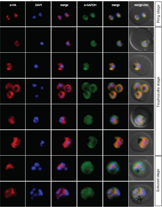
Localisation of PF11_0293 during the IDC. Localisation of the tagged protein was visualised using anti-HA antibodies (red). Antibodies against GAPDH were used to visualise the cytosolic compartment. DAPI was used to visualise the nucleus. DIC images are shown as reference. The protein is localized outside but in close proximity to the DAPI signal. These patterns were clearly distinct from the cytosolic GAPDH signal suggesting that it may be associated with the nuclear periphery. In addition, the protein appeared to accumulate in the cytosol in mature trophozoites.Oehring SC, Woodcroft BJ, Moes S, Wetzel J, Dietz O, Pulfer A, Dekiwadia C, Maeser P, Flueck C, Witmer K, Brancucci NM, Niederwieser I, Jenoe P, Ralph SA, Voss TS. Organellar proteomics reveals hundreds of novel nuclear proteins in the malaria parasite Plasmodium falciparum. Genome Biol. 2012 13(11):R108
See original on MMP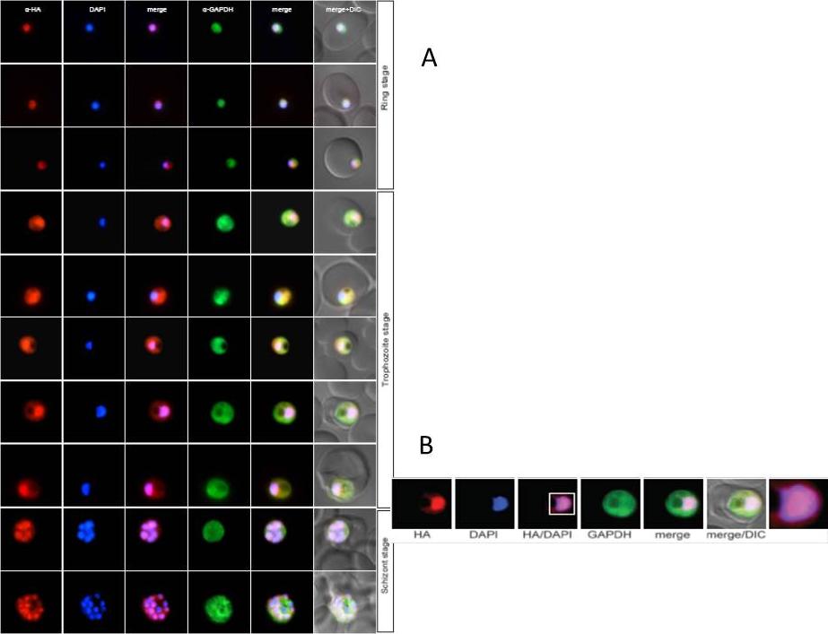
Localisation of PF11_0332 during the IDC. Localisation of the tagged protein was visualised using anti-HA antibodies (red). Antibodies against GAPDH were used to visualise the cytosolic compartment. DAPI was used to visualise the nucleus. DIC images are shown as reference. The protein shows a diffuse pattern in the nucleus.Oehring SC, Woodcroft BJ, Moes S, Wetzel J, Dietz O, Pulfer A, Dekiwadia C, Maeser P, Flueck C, Witmer K, Brancucci NM, Niederwieser I, Jenoe P, Ralph SA, Voss TS. Organellar proteomics reveals hundreds of novel nuclear proteins in the malaria parasite Plasmodium falciparum. Genome Biol. 2012 Nov 26;13(11):R108.
See original on MMP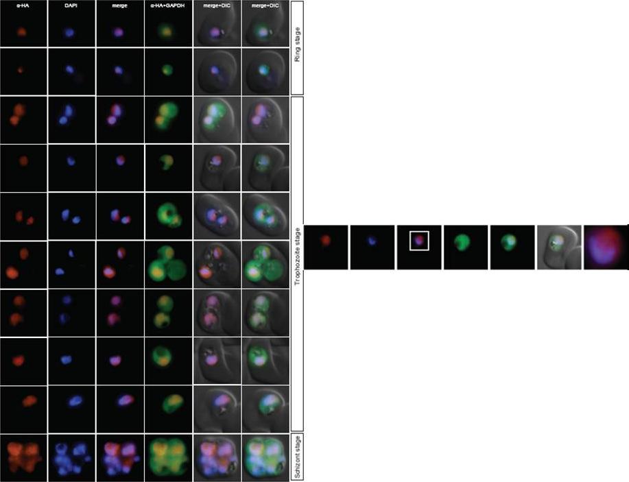
Localisation of PF13_0042 during the IDC. Localisation of the tagged protein was visualised using anti-HA antibodies (red). Antibodies against GAPDH were used to visualise the cytosolic compartment. DAPI was used to visualise the nucleus. DIC images are shown as reference. The protein is associated with the nucleus.Oehring SC, Woodcroft BJ, Moes S, Wetzel J, Dietz O, Pulfer A, Dekiwadia C, Maeser P, Flueck C, Witmer K, Brancucci NM, Niederwieser I, Jenoe P, Ralph SA, Voss TS. Organellar proteomics reveals hundreds of novel nuclear proteins in the malaria parasite Plasmodium falciparum. Genome Biol. 2012 Nov 26;13(11):R108.
See original on MMP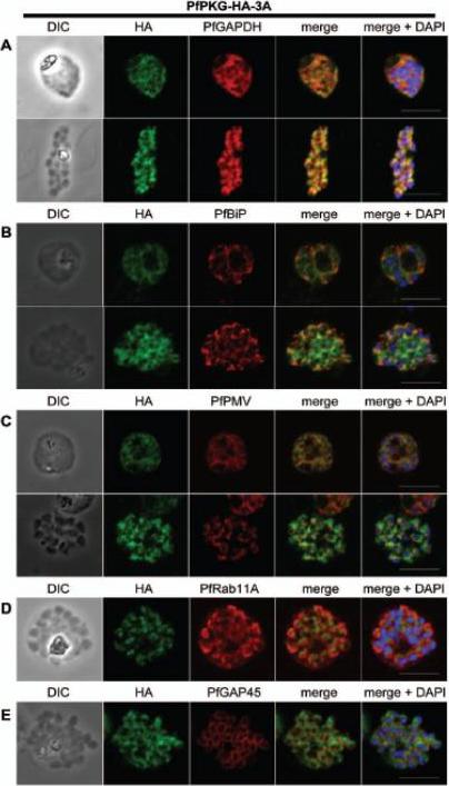
Subcellular location of PfPKG in mature schizonts. Dual immunofluorescent detection of PfPKG-HA in fixed smears of early and late schizonts of the PfPKG-HA-3A clone together with (A) GAPDH; (B) BiP; (C) PMV; (D) Rab11A and (E) GAP45. Representative images are shown for each antibody, together with bright field images (first column) and parasite nuclei stained with DAPI (in the merged image). Bars ,5 mm. Significant overlap between the PKG-HA and GAPDH in early and late blood stage schizonts (A), both are in the cytosol. PfPKG-HA also partially overlapped with that of the ER membrane protein PMV and that of the ER lumen marker PfBiP (B and C) in both early and late schizonts. Rab11A is thought to be involved in vesicle trafficking and localises to the rhoptries and the inner membrane complex (IMC) at the apical end of the merozoite. In mature schizonts, the location of PKG-HA appeared to be largely distinct from that of Rab11A (D). PKG-HA appeared to be absent from the IMC in segmented schizonts, since it did not colocalise with the IMC marker, the glideosome associated protein 45. Hopp CS, Flueck C, Solyakov L, Tobin A, Baker DA. Spatiotemporal and Functional Characterisation of the Plasmodium falciparum cGMP-Dependent Protein Kinase. PLoS One. 2012;7(11):e48206.
See original on MMP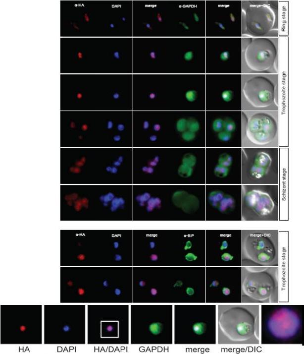
Localisation of PF14_0393 during the IDC. Localisation of the tagged protein was visualised using anti-HA antibodies (red). Antibodies against GAPDH were used to visualise the cytosolic compartment. Antibodies against PfBIP were used to visualise the ER (bottom). DAPI was used to visualise the nucleus. DIC images are shown as reference. The protein displayed a condensed appearance in the nucleus throughout the IDC. Oehring SC, Woodcroft BJ, Moes S, Wetzel J, Dietz O, Pulfer A, Dekiwadia C, Maeser P, Flueck C, Witmer K, Brancucci NM, Niederwieser I, Jenoe P, Ralph SA, Voss TS. Organellar proteomics reveals hundreds of novel nuclear proteins in the malaria parasite Plasmodium falciparum. Genome Biol. 2012 13(11):R108.
See original on MMP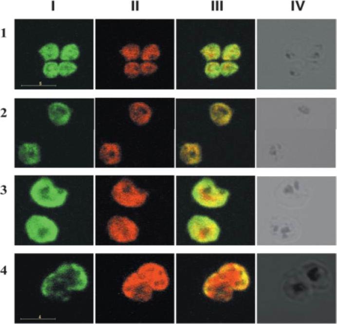
IFA of merozoites stained with a mAb specific for pfGAPDH, pfAldolase and RAP-1. Row 1 demonstrates merozoites stained with anti-pfGAPDH mAb (green, column I), anti-pfAldolase mAb (red, column II) and the overlay of both images (column III). Row 2 shows merozoites stained with anti-pfGAPDH mAb (green, column I) and anti-RAP-1 mAb 5-2 (red, column II) and the overlay of both images (column III). pfAldolase and pfGAPDH were colocalized in the early stages of parasite development since both, the green and the red signals, were super-imposed resulting in orange signals. At 40 h post synchronization the green signal of the anti-pfGAPDH staining was largely confined to the periphery of the schizont, while the red signal of the anti-pfAldolase staining remained distributed equally throughout the schizont. pfGAPDH and pfAldolase were preferentially localized at opposite cell poles. pfGAPDH was preferentially co-localized with RAP-1 which is associated with the apical complex of the merozoites.Daubenberger CA, Tisdale EJ, Curcic M, Diaz D, Silvie O, Mazier D, Eling W, Bohrmann B, Matile H, Pluschke G. The N'-terminal domain of glyceraldehyde-3-phosphate dehydrogenase of the apicomplexan Plasmodium falciparum mediates GTPase Rab2-dependent recruitment to membranes. Biol Chem. 2003 384:1227-37. Copyright Walter de Gruyter GmbH 2009
See original on MMP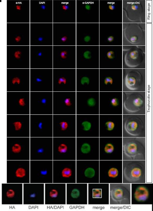
Localisation of PFC0126c during the IDC. Localisation of the tagged protein was visualised using anti-HA antibodies (red). Antibodies against GAPDH were used to visualise the cytosolic compartment. DAPI was used to visualise the nucleus. DIC images are shown as reference. The protein is localized to a cytoplasmic structure distinct from the cytosolic and ER compartments, which likely represents the mitochondrion.Oehring SC, Woodcroft BJ, Moes S, Wetzel J, Dietz O, Pulfer A, Dekiwadia C, Maeser P, Flueck C, Witmer K, Brancucci NM, Niederwieser I, Jenoe P, Ralph SA, Voss TS. Organellar proteomics reveals hundreds of novel nuclear proteins in the malaria parasite Plasmodium falciparum. Genome Biol. 2012 Nov 26;13(11):R108.
See original on MMP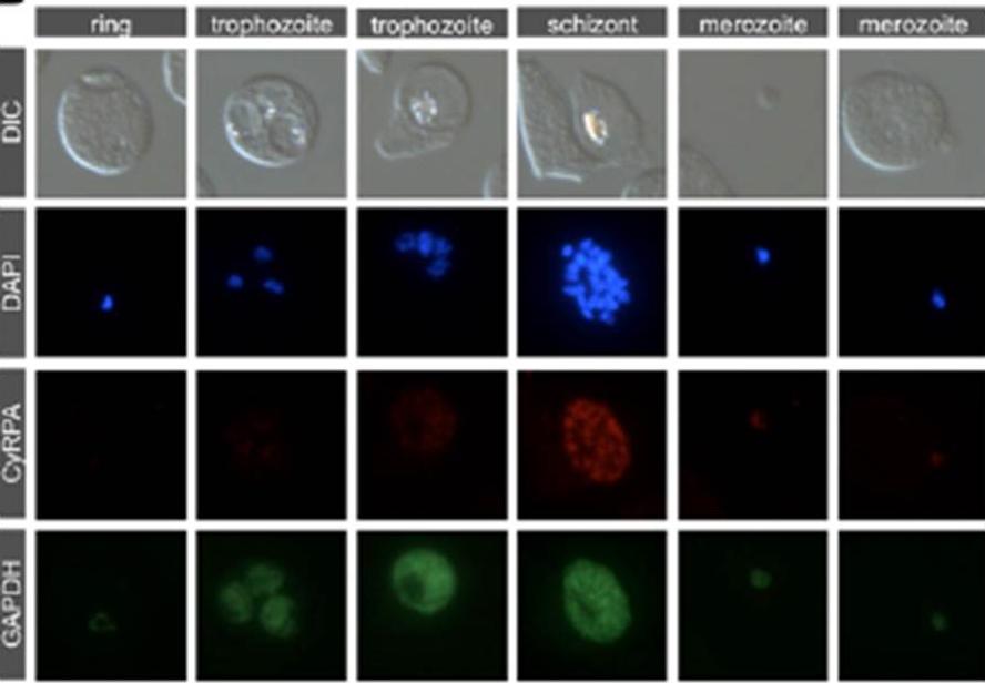
Stage-specific expression of CyRPA in late asexual blood-stage parasites. Indirect immunofluorescence stainings of asexual bloodstage parasites confirmed stage-specific expression in schizont stages and free merozoites. Methanol/acetone fixed P. falciparum 3D7 parasites were probed with anti-CyRPA mAb c06 (red) and anti-GAPDH mAb (green). Exposure times were identical for all pictures of the same channel. Nuclei were stained with DAPI (blue). Original magnification x1008.Dreyer AM, Matile H, Papastogiannidis P, Kamber J, Favuzza P, Voss TS, Wittlin S, Pluschke G. Passive immunoprotection of Plasmodium falciparum-infected mice designates the CyRPA as candidate malaria vaccine antigen. J Immunol. 2012 188(12):6225-37.
See original on MMP
Localization of CyRPA to the merozoite apex by immunofluorescence staining. P. falciparum 3D7 merozoites (A) or schizont stages (B) were coimmunostained with anti-CyRPA mAb c06 (red) and anti-GAPDH Abs (marker for cytosol), AMA-1 (marker for micronemes), RAP-1 (marker for rhoptry bulbs), MSP-1 (marker for merozoite surface), or MSP-5 (green). Nuclei were stained with DAPI (blue). Original magnification x1008. CyRPA is localized at the merozoite apex.Dreyer AM, Matile H, Papastogiannidis P, Kamber J, Favuzza P, Voss TS, Wittlin S, Pluschke G. Passive immunoprotection of Plasmodium falciparum-infected mice designates the CyRPA as candidate malaria vaccine antigen. J Immunol. 2012 188(12):6225-37.
See original on MMP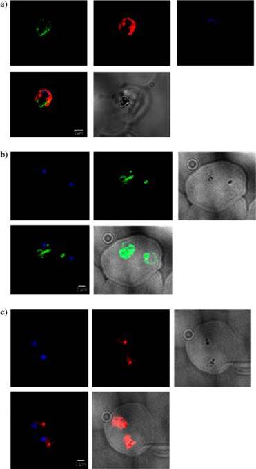
Immunofluorescence studies with P. falciparum trophozoites that were treated with 10 μg/mL OZ277 for 2 h. (a) In immunofluorescence colocalization studies, the blood smears were fixed with 5% formaldehyde and 0.01% glutaraldehyde, permeabilized with 0.5% Triton-X-100, and blocked with 1% BSA in PBS for 1 h. The primary antibodies used were directly labeled OZH04-2/2 (Alexa 488, green signal) and GAPDH (Alexa 594, red signal). For both, the exposure time was 2 s. Nuclei were stained with DAPI (blue signal, exposure time of 0.05 s). The bottom row left shows the merge of OZH04-2/2, GAPDH, and DAPI and the bottom row right, the reference image (exposure of 1 s). Scale bar = 2 μm. (b) Same as (a), except that unlabeled primary antibodies OZH04-2/2 were used. After six washes with PBS, 20 μg/mL of the secondary antibody goat antimouse Alexa 488 (green signal) was incubated for 1 h at room temperature. The exposure time was 0.1 s. Nuclei were stained with DAPI (blue signal, exposure time of 0.05 s). The bottom row left shows the merge of OZH04-2/2 and DAPI, and the bottom row right shows the merge of OZH04-2/2 and the reference image (exposure of 1 s). Scale bar = 2 μm. (c) Same as (b), except that the secondary antibody used was goat antimouse Alexa 594 with an exposure of 1 s (red signal).Jourdan J, Matile H, Reift E, Biehlmaier O, Dong Y, Wang X, Mäser P, Vennerstrom JL, Wittlin S. Monoclonal Antibodies That Recognize the Alkylation Signature of Antimalarial Ozonides OZ277 (Arterolane) and OZ439 (Artefenomel). ACS Infect Dis. 2016 Jan 8;2(1):54-61.
See original on MMP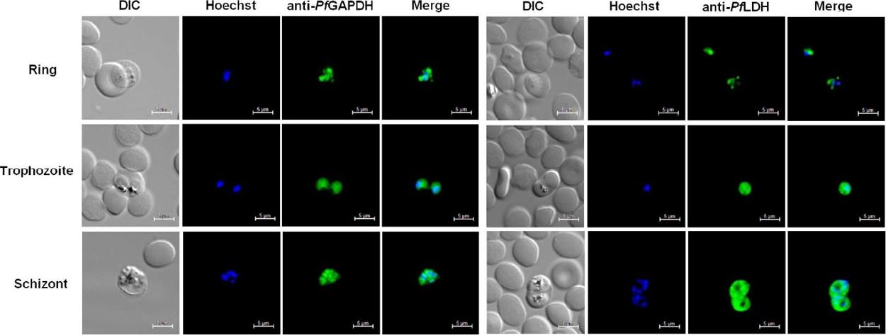
Immunolocalization of P. falciparum LDH and GAPDH in asexual P. falciparum (3D7) parasites grown in vitro, synchronized by sorbitol treatment and prepared at ring, trophozoite and schizont stages as indicated alongside the panels. Fixed parasites were probed with chicken anti-rPfLDH or anti-rPfGAPDH antibodies and the chicken antibodies were detected with a Cy3-conjugated antibody. Parasite DNA was stained with Hoechst and the scale bar represents 5 μm. DIC images are shown as reference. PfGAPDH and PfLDH showed very similar 442 distribution throughout the cytoplasm of the parasites, however PfGAPDH seemed to localize to the nucleus in schizont stages.Krause RGE, Hurdayal R, Choveaux D, Przyborski J, Coetzer THT, Goldring JPD. Plasmodium glyceraldehyde-3-phosphate dehydrogenase: A potential malaria diagnostic target. Exp Parasitol. 2017 May 25. [Epub ahead of print] PMID: 28552792
See original on MMPMore information
| PlasmoDB | PF3D7_1462800 |
| GeneDB | PF3D7_1462800 |
| Malaria Metabolic Pathways | Localisation images Pathways mapped to |
| Previous ID(s) | PF14_0598 |
| Orthologs | PBANKA_1326400 , PCHAS_1329700 , PKNH_1218700 , PVP01_1244000 , PVX_117322 , PY17X_1330200 |
| Google Scholar | Search for all mentions of this gene |