PF3D7_1452300 DER1-like protein (Der1-1)
Disruptability [+]
| Species | Disruptability | Reference | Submitter | |
|---|---|---|---|---|
| P. berghei ANKA |
Possible |
PlasmoGEM (Barseq) | PlasmoGEM | |
Mutant phenotypes [+]
| Species | Stage | Phenotype | Reference | Submitter |
|---|---|---|---|---|
| P. berghei ANKA | Asexual |
Attenuated |
PlasmoGEM (Barseq) | PlasmoGEM |
Imaging data (from Malaria Metabolic Pathways)
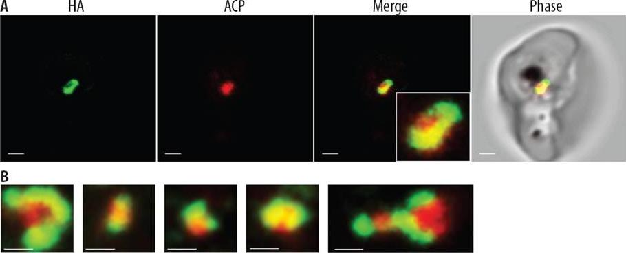
PfsDer1-1-HA is located at the periphery of the apicoplast. (A) Confocal images of chemically fixed PfsDer1-1-HA parasites labelled with anti-HA (green) and anti-ACP antibodies (red), showing that PfsDer1-1-HA is associated with ACP (merge), but is peripheral, similar to PfTic22-HA. Inset shows a 3.3x zoom of the localizations. (B) More examples of peripheral localization, similar to the inset above. Scale bar shows 0.5 mm.Kalanon M, Tonkin CJ, McFadden GI. Characterisation of two putative protein translocation components in the apicoplast of Plasmodium falciparum. Eukaryot Cell. 2009 8(8):1146-54
See original on MMP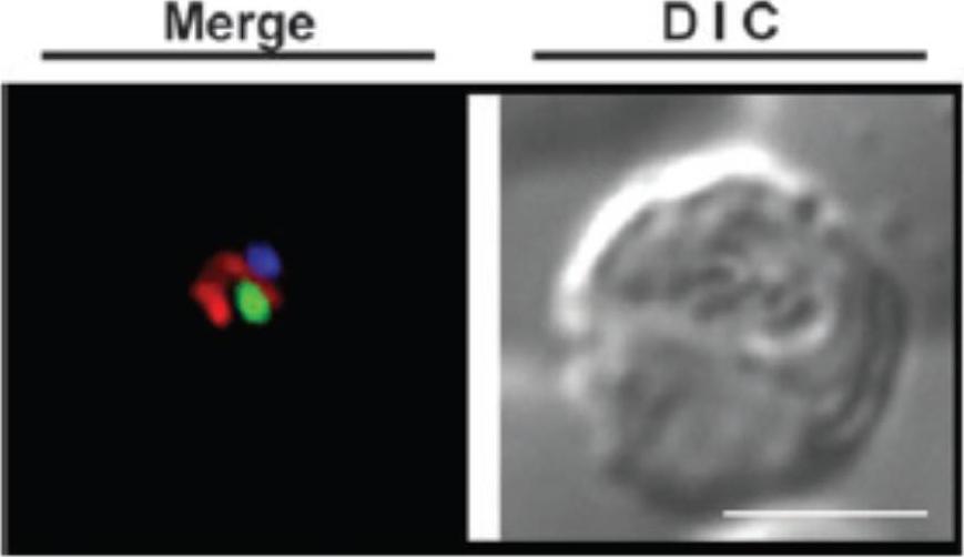
In vivo localization of the Plasmodium falciparum Localization of the symbiont-specific Der1-1 BTS–GFP fusion protein in P. falciparum 3D7 blood stage parasites (3D7sDer1-1 BTSGFP ; in green) additionally stained with MitoTracker (in red) and Hoechst (in blue). The typical ‘‘dot’’ pattern of apicoplast targeted GFP is clearly evident. Scale bar represents 4 mm. The BTS of the P. falciparum symbiont Der1-1 (3D7sDer1-1 BTSGFP) is able to direct GFP to the apicoplast BTS - bipartite topogenic signal, comprised of an N-terminal signal peptide and a plastid transit peptide, the sequence of which determines whether 2 membranes (into the PPC) or 4 membranes (into the plastid stroma) are crossed. Staining with Hoechst and MitoTracker proves that the GFP expression does not display a nuclear or mitochondrial localization.Sommer MS, Gould SB, Lehmann P, Gruber A, Przyborski JM, Maier UG. Der1-mediated preprotein import into the periplastid compartment of chromalveolates? Mol Biol Evol. 2007 24(4):918-28
See original on MMP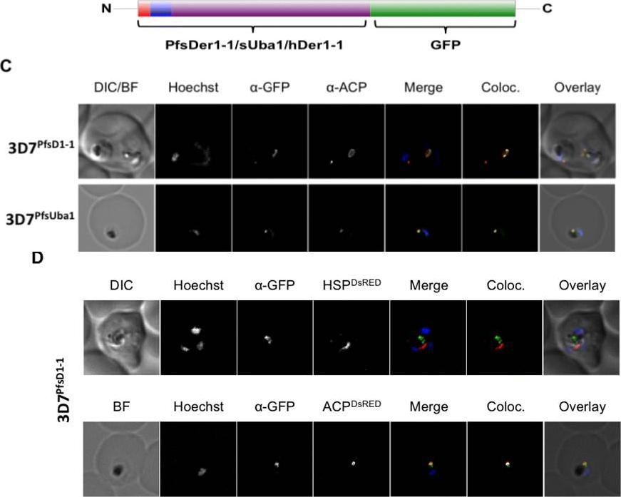
C. In both 3D7PfsD1-1 and 3D7PfsUba1 lines, GFP and ACP signals colocalise, suggesting that both are resident apicoplast proteins. Co-transfected 3D7sD1-1 with a construct encoding either the BTS of the apicoplast resident protein ACP or the mitochondrial targeting sequence of mitochondrial PfHsp60, fused to the red fluorescent protein DsRed. D. Fixed cells co-labelled with anti-GFP antibodies, ACP-DsRed PFB0385w and GFP signals co-localise, whilst HSP60-DsRed and GFP signal do not. Thus, the full-length PfsDer1-1-GFP fusion protein is transported to the apicoplast.Spork S, Hiss JA, Mandel K, Sommer M, Kooij TW, Chu T, Schneider G, Maier UG, Przyborski JM. An unusual ERAD-like complex is targeted to the apicoplast of Plasmodium falciparum. Eukaryot Cell. 2009 ;8(8):1134-45
See original on MMP
(A) Live cell imaging of co-transfectants expressing saGFP (self-assembling split GFP) fragments in various cellular compartments. In merge and overlay: green, GFP; blue, Hoechst. (B) Vectors available for analysis of cellular compartmentalisation using saGFP. Numbers in targeting sequence refer to N-terminal amino acids. B, Blasticidin-S-deaminase resistance cassette; D, hDHFR resistance cassette. *Can be used to generate fusions as multiple cloning sites situated in front of saGFP coding sequence. Külzer S, Petersen W, Baser A, Mandel K, Przyborski JM. Use of self-assembling GFP to determine protein topology and compartmentalisation in the Plasmodium falciparum-infected erythrocyte. Mol Biochem Parasitol. 2012 187(2):87-90.
See original on MMPMore information
| PlasmoDB | PF3D7_1452300 |
| GeneDB | PF3D7_1452300 |
| Malaria Metabolic Pathways | Localisation images Pathways mapped to |
| Previous ID(s) | PF14_0498 |
| Orthologs | PBANKA_1316000 , PCHAS_1319300 , PKNH_1229800 , PVP01_1254700 , PVX_117865 , PY17X_1319800 |
| Google Scholar | Search for all mentions of this gene |