PF3D7_1448400 ubiquitin-protein ligase, putative (HRD3)
Disruptability [+]
| Species | Disruptability | Reference | Submitter |
|---|---|---|---|
| P. falciparum 3D7 |
Possible |
USF piggyBac screen (Insert. mut.) | USF PiggyBac Screen |
Mutant phenotypes [+]
None reported yet. Please press the '+' button above to add one.Imaging data (from Malaria Metabolic Pathways)
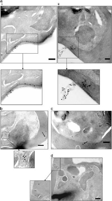
Ultrathin sections of P. falciparum-infected erythrocytes (at late trophozoite/schizont stages) were labeled with specific sera (against the four selected proteins) and gold-conjugated secondary antibody. Localization is depicted as black dots (of gold particles) for PfSEL1 (PFB0190c) - (a, i and ii), PfSEL2 = Hrd3 (b), PfEK (c), and PfEP (d). Enlarged panels and arrows show detailed images of the intracellular staining pattern. Scale bar, 250 nm. PfSEL1 showed a secreted/ extra-cellular staining pattern, localizing in the iRBC cytosol close to the erythrocyte plasma membrane. The gold particle staining showed the protein coating the surface of the iRBC and also being released out in the extra-cellular milieu. PfSEL2 (seen as dots coating vesicle-like structures) and PfEK also showed localization close to iRBC plasma membrane while being exported out of the infected red cells (b and c). PfEP localized in the extensive membranous network of the parasite from where it seemed to be trafficked to the iRBC cytosol and plasma membrane (d). Singh M, Mukherjee P, Narayanasamy K, Arora R, Sen SD, Gupta S, Natarajan K, Malhotra P. Proteome analysis of Plasmodium falciparum extracellular secretory antigens at asexual blood stages reveals a cohort of proteins with possible roles in immune modulation and signaling. Mol Cell Proteomics. 2009 8(9):2102-18.
See original on MMP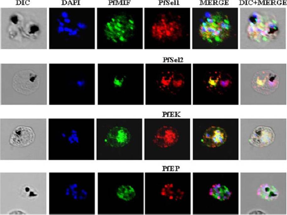
Indirect immunofluorescence assay and confocal microscopy to localize the selected excretory-secretory antigens by coimmunostaining of P. falciparum-infected erythrocytes. Air-dried infected erythrocytes were incubated with mouse antibodies specific to the selected proteins at 1:100 dilution (column 4; red). Rabbit aPfMIF antibody was used as a co-localization marker (column 3; green). The parasite nuclei were stained with 4’,6-diamidino-2-phenylindole (DAPI) (column 2; blue). Columns 1, 5, and 6 show a differential interference contrast (DIC) image, merged image, and differential interference contrast with merged image, respectively. Images showed punctate vesicle-like staining of the infected RBCs with antibodies to PfSEL1 (PFB0190c), PfSEL2 (Hrd3), PfEKand PfEP. PfEK also showing specific rimlike staining of the plasma membrane of the infected RBC. PfMIF is exported via the Maurer clefts to the extracellular medium after schizont rupture. A partial co-localization of PfMIF was seen with each of the four proteins, suggesting a distinct pathway for the release of these ESAs.Singh M, Mukherjee P, Narayanasamy K, Arora R, Sen SD, Gupta S, Natarajan K, Malhotra P. Proteome analysis of Plasmodium falciparum extracellular secretory antigens at asexual blood stages reveals a cohort of proteins with possible roles in immune modulation and signaling. Mol Cell Proteomics. 2009 8(9):2102-18.
See original on MMPMore information
| PlasmoDB | PF3D7_1448400 |
| GeneDB | PF3D7_1448400 |
| Malaria Metabolic Pathways | Localisation images Pathways mapped to |
| Previous ID(s) | PF14_0462 |
| Orthologs | PBANKA_1312200 , PCHAS_1315500 , PKNH_1233700 , PVP01_1258600 , PVX_118065 , PY17X_1316000 |
| Google Scholar | Search for all mentions of this gene |