PF3D7_1446200 M17 leucyl aminopeptidase (LAP)
Disruptability [+]
| Species | Disruptability | Reference | Submitter | |
|---|---|---|---|---|
| P. falciparum 3D7 |
Refractory |
USF piggyBac screen (Insert. mut.) | USF PiggyBac Screen | |
| P. berghei ANKA |
Possible |
RMgm-814 | Imported from RMgmDB | |
Mutant phenotypes [+]
| Species | Stage | Phenotype | Reference | Submitter |
|---|---|---|---|---|
| P. berghei ANKA | Asexual |
Difference from wild-type |
RMgm-814
Strongly reduced growth rate of blood stages (normal levels of hemozin production) |
Imported from RMgmDB |
| P. berghei ANKA | Asexual |
Attenuated |
PlasmoGEM (Barseq) | PlasmoGEM |
Imaging data (from Malaria Metabolic Pathways)
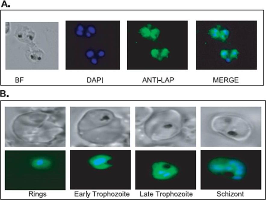
Localization of the P. falciparum M17 leucyl aminopeptidase in the intra-erythrocytic parasites. A, immunofluorescence assays were carried out using air-dried P. falciparum-infected red blood cells fixed with acetone. Cells were probed with mouse anti-M17 peptide (1/250) and then Cy2-conjugated goat anti-mouse antibodies. B, transgenic P. falciparum strain D10 parasites expressing the complete M17 leucyl aminopeptidase linked to green fluorescent protein were visualized in a live fluorescence assay. The parasite nuclei were visualized with Hoechst dye (0.5 mg/ml). Samples were viewed on an Axioscope 2 Mot + (Zeiss) equipped with a Zeiss 63x/1.4 Plan Apochromat lens. Images were captured with an Axiocam MRm camera (Zeiss) using Axiovision AC software (Zeiss). BF, bright field; DAPI, 4,6-diamidino-2-phenylindole.Stack CM, Lowther J, Cunningham E, Donnelly S, Gardiner DL, Trenholme KR, Skinner-Adams TS, Teuscher F, Grembecka J, Mucha A, Kafarski P, Lua L, Bell A, Dalton JP. Characterization of the Plasmodium falciparum M17 leucyl aminopeptidase. A protease involved in amino acid regulation with potential for antimalarial drug development. J Biol Chem. 2007 282:2069-2080.
See original on MMP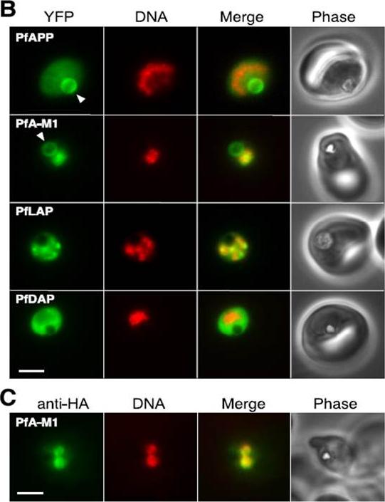
Localization of aminopeptidases in P. falciparum blood stages.PfAPP Aminopeptidase P; PfA-M1 Aminopeptidase N; PfLAP Leucyl aminopeptidase; PfDAP Aspartyl aminopeptidase B, Epifluorescence microscopy of live parasites expressing fusions of YFP with the indicated aminopeptidases. YFP fluorescence is pseudocolored green and Hoechst 33342 (DNA) fluorescence is pseudocolored red. The arrowhead indicates the food vacuole. Bar, 2 mm. C, indirect immunofluorescence detection of PfA-M1-HA. Secondary antibody fluorescence is pseudocolored green and DAPI (DNA) fluorescence is pseudocolored red. Bar, 2 mm.Dalal S, Klemba M. Roles for two aminopeptidases in vacuolar hemoglobin catabolism in Plasmodium falciparum. J Biol Chem. 2007 282:35978-87.
See original on MMPMore information
| PlasmoDB | PF3D7_1446200 |
| GeneDB | PF3D7_1446200 |
| Malaria Metabolic Pathways | Localisation images Pathways mapped to |
| Previous ID(s) | PF14_0439 |
| Orthologs | PBANKA_1309900 , PCHAS_1313100 , PCHAS_1313400 , PCHAS_1313500 , PKNH_1236000 , PVP01_1260800 , PVX_118180 , PY17X_1313800 |
| Google Scholar | Search for all mentions of this gene |