PF3D7_1444800 fructose-bisphosphate aldolase (FBPA)
Disruptability [+]
| Species | Disruptability | Reference | Submitter | |
|---|---|---|---|---|
| P. falciparum 3D7 |
Refractory |
USF piggyBac screen (Insert. mut.) | USF PiggyBac Screen | |
| P. berghei ANKA |
Refractory |
PlasmoGEM (Barseq) | PlasmoGEM | |
Mutant phenotypes [+]
None reported yet. Please press the '+' button above to add one.Imaging data (from Malaria Metabolic Pathways)
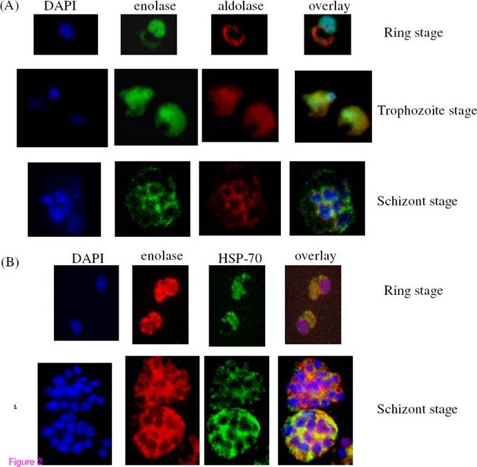
Immunofluorescence assays for the localization of enolase, aldolase and HSP-70 in P. falciparum asexual stages (ring, trophozoite and schizont). (A) P. falciparum infected red blood cells were treated with DAPI (blue), mouse anti rPfen antibody (green), rabbit anti-P. falciparum aldolase antibody (red). (B) Cells were treated with DAPI, rabbit rPfen antibody (red), and mouse anti Pf HSP-70 antibody (green). Overlay panels show the merged of the three images.Pal Bhowmick I, Kumar N, Sharma S, Coppens I, Jarori GK. Plasmodium falciparum enolase: stage-specific expression and sub-cellular localization. Malar J. 2009 8(1):179
See original on MMP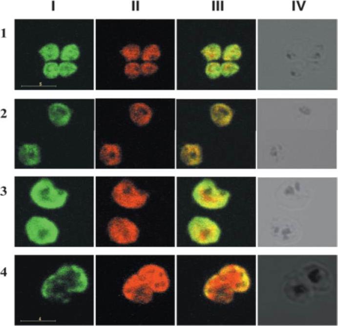
IFA of merozoites stained with a mAb specific for pfGAPDH, pfAldolase and RAP-1. Row 1 demonstrates merozoites stained with anti-pfGAPDH mAb (green, column I), anti-pfAldolase mAb (red, column II) and the overlay of both images (column III). Row 2 shows merozoites stained with anti-pfGAPDH mAb (green, column I) and anti-RAP-1 mAb 5-2 (red, column II) and the overlay of both images (column III). pfAldolase and pfGAPDH were colocalized in the early stages of parasite development since both, the green and the red signals, were super-imposed resulting in orange signals. At 40 h post synchronization the green signal of the anti-pfGAPDH staining was largely confined to the periphery of the schizont, while the red signal of the anti-pfAldolase staining remained distributed equally throughout the schizont. pfGAPDH and pfAldolase were preferentially localized at opposite cell poles. pfGAPDH was preferentially co-localized with RAP-1 which is associated with the apical complex of the merozoites.Daubenberger CA, Tisdale EJ, Curcic M, Diaz D, Silvie O, Mazier D, Eling W, Bohrmann B, Matile H, Pluschke G. The N'-terminal domain of glyceraldehyde-3-phosphate dehydrogenase of the apicomplexan Plasmodium falciparum mediates GTPase Rab2-dependent recruitment to membranes. Biol Chem. 2003 384:1227-37. Copyright Walter de Gruyter GmbH 2009
See original on MMP
Longitudinal intensity profiling localises actin and aldolase towards the rear of the invading merozoite’s tight junction. Single-slice images of example merozoites for the longitudinal intensity profiling of mRON4 (green) vs (left panel) rActin (rAct) (red) or (right panel) rAlodlase (rAldo) (red). Scale bars = 1 μm. Red boxes in DIC images indicate zoomed regions for middle and right panels. Arrows indicate the position of the RON4 labelled tight junctionRiglar DT, Whitehead L, Cowman AF, Rogers KL, Baum J. Localization-based imaging of malarial antigens during red cell entry reaffirms role for AMA1 but not MTRAP in invasion. J Cell Sci. 2015. [Epub ahead of print] PMID:
See original on MMPMore information
| PlasmoDB | PF3D7_1444800 |
| GeneDB | PF3D7_1444800 |
| Malaria Metabolic Pathways | Localisation images Pathways mapped to |
| Previous ID(s) | PF14_0425 |
| Orthologs | PBANKA_1308600 , PCHAS_1311800 , PKNH_1237500 , PVP01_1262200 , PVX_118255 , PY17X_1312400 |
| Google Scholar | Search for all mentions of this gene |