PF3D7_1440300 porphobilinogen synthase (PBGS)
Disruptability [+]
| Species | Disruptability | Reference | Submitter | |
|---|---|---|---|---|
| P. falciparum 3D7 |
Refractory |
USF piggyBac screen (Insert. mut.) | USF PiggyBac Screen | |
| P. berghei ANKA |
Refractory |
PlasmoGEM (Barseq) | PlasmoGEM | |
Mutant phenotypes [+]
None reported yet. Please press the '+' button above to add one.Imaging data (from Malaria Metabolic Pathways)
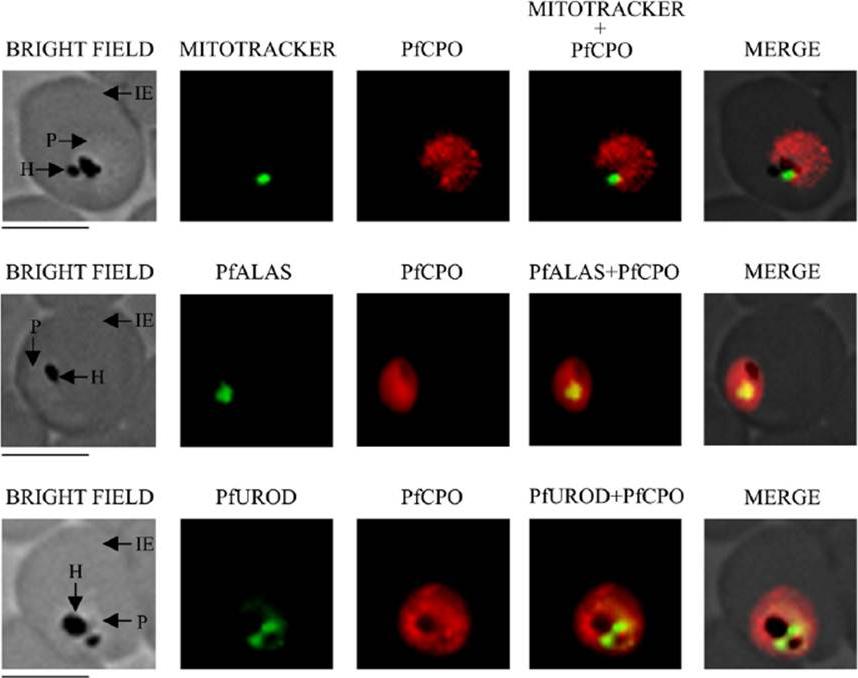
Localization of PfCPO by immunofluorescence microscopy. Localization of PfCPO with Mitotracker green (mitochondrial marker), PfALAS (mitochondrial marker) and PfUROD (apicoplast marker) was studied. IE, infected erythrocyte; P, parasite; H, hemozoin. Scale bar=5 μm. results presented in Fig. 4 indicate that the PfCPO signal was distributed throughout the parasite, but did not colocalize with that of Mitotracker dye (mitochondrial marker), PfALAS (mitochondrial marker) or PfUROD (apicoplast marker).Nagaraj VA, Prasad D, Arumugam R, Rangarajan PN, Padmanaban G. Characterization of coproporphyrinogen III oxidase in Plasmodium falciparum cytosol. Parasitol Int. 2010 59:121-127. Copyright Elsevier 2011
See original on MMP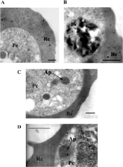
Localization of PfALAD in P. falciparum by immunoelectron microscopy. A and B, localization of host ALAD in the parasite and red blood cell. C and D, localization of PfALAD in the apicoplast with multimembrane structure. The abundant presence of host ALAD in the cytoplasm and food vacuole and the sparse occurrence of PfALAD in the apicoplast (shown by the arrow) of P. falciparum is indicated by the distribution of gold particles. Scale bar 0.5 mm. Pc, parasite cytoplasm. Rc, red cell cytoplasm. Fv, food vacuole. Ap, apicoplast. The enzyme has been localized to the apicoplast of the malaria parasite.Dhanasekaran S, Chandra NR, Chandrasekhar Sagar BK, Rangarajan PN, Padmanaban G. Delta-aminolevulinic acid dehydratase from Plasmodium falciparum: indigenous versus imported. J Biol Chem. 2004 279(8):6934-42
See original on MMP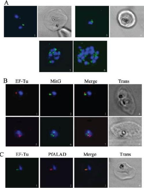
EF-Tu is localized within the apicoplast of P. falciparum. A. Fluorescence (green) localizes within the apicoplast in the middle trophozoite (1), late trophozoite (3) and schizont (5 and 6) stages. Images 2 and 4 are the corresponding phase contrast scans of 1 and 3 respectively. Green fluorescence is associated with most nuclei (blue) in images 5 and 6 (all apicoplasts may not lie in the photographed focal plane). B. Closely associated but distinct fluorescence for apicoplast (red) and mitochondria (green) is seen in the middle trophozoite (1–4) and early schizont stages (5–8). C. Overlapping signals are observed for EF-Tu (green) and PfALAD (red) in the middle trophozoite stage (1–4).Chaubey S, Kumar A, Singh D, Habib S. The apicoplast of Plasmodium falciparum is translationally active. Mol Microbiol. 2005 56:81-9. Copyright John Wiley & Sons Ltd. 2010
See original on MMP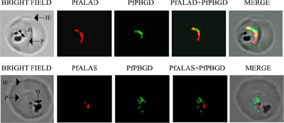
Localization of porphobilinogen deaminase PfPBGD with d-aminolevulinate dehydratase PfALAD and d-aminolevulinic acid synthase PfALAS in P. falciparum by immunofluorescence. PGDB and PfALAD are localized to the apicoplast. PfALAS is localized to the mitochondrion. IE, infected erythrocyte; P, parasite; H, hemozoin.Sato S, Clough B, Coates L, Wilson RJ. Enzymes for heme biosynthesis are found in both the mitochondrion and plastid of the malaria parasite Plasmodium falciparum. Protist. 2004 155:117-25. Copyright Elsevier 2009.
See original on MMP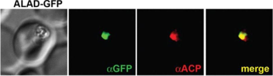
IFM images of fixed 3D7 parasites expressing full-length ALAD tagged at its endogenous locus with C-terminal GFP confirm targeting to the parasite apicoplast.Parasites were stained with αGFP and αACP (ACP, apicoplast marker).Sigala PA, Crowley JR, Henderson JP, Goldberg DE. Deconvoluting heme biosynthesis to target blood-stage malaria parasites. Elife. 2015 Jul 14;4.
See original on MMPMore information
| PlasmoDB | PF3D7_1440300 |
| GeneDB | PF3D7_1440300 |
| Malaria Metabolic Pathways | Localisation images Pathways mapped to |
| Previous ID(s) | PF14_0381 |
| Orthologs | PBANKA_1304100 , PCHAS_1307300 , PKNH_1242100 , PVP01_1266800 , PVX_118480 , PY17X_1307900 , PY17X_1308000 |
| Google Scholar | Search for all mentions of this gene |