PF3D7_1438900 thioredoxin peroxidase 1 (Trx-Px1)
Disruptability [+]
| Species | Disruptability | Reference | Submitter | |
|---|---|---|---|---|
| P. falciparum 3D7 |
Possible |
12860402 hypersensitive to stress |
Theo Sanderson, Wellcome Trust Sanger Institute | |
| P. falciparum 3D7 |
Possible |
USF piggyBac screen (Insert. mut.) | USF PiggyBac Screen | |
| P. berghei ANKA |
Possible |
RMgm-206 | Imported from RMgmDB | |
| P. berghei ANKA |
Possible |
PlasmoGEM (Barseq) | PlasmoGEM | |
Mutant phenotypes [+]
| Species | Stage | Phenotype | Reference | Submitter |
|---|---|---|---|---|
| P. falciparum 3D7 | Asexual |
No difference |
12860402 hypersensitive to stress |
Theo Sanderson, Wellcome Trust Sanger Institute |
| P. falciparum 3D7 | Asexual |
Transcriptional effect |
35939693 (Knock down)
\'Furthermore, we show that PfTPx-1 knockdown alters the var switch rate as well as activation of additional gene subsets.\' |
Theo Sanderson, Francis Crick Institute |
| P. berghei ANKA | Asexual |
No difference |
RMgm-206 | Imported from RMgmDB |
| P. berghei ANKA | Asexual |
No difference |
PlasmoGEM (Barseq) | PlasmoGEM |
| P. berghei ANKA | Gametocyte |
Difference from wild-type |
RMgm-206
Reduced production of gametocytes (60% fewer gametocytes compared to wild type) |
Imported from RMgmDB |
| P. berghei ANKA | Oocyst |
No difference |
RMgm-206 | Imported from RMgmDB |
| P. berghei ANKA | Sporozoite |
Difference from wild-type |
RMgm-206
Normal numbers of oocyst are produced. The number of mature oocysts containing sporozoites and the number of salivary gland sporozoites is reduced compared to wild type. Infectivity of salivary gland sporozoites is reduced as shown by a prolonged pre-patent period after intravenous inoculation of sporozoites. |
Imported from RMgmDB |
| P. berghei ANKA | Liver |
Difference from wild-type |
RMgm-206
Infectivity of salivary gland sporozoites is reduced as shown by a prolonged pre-patent period after intravenous inoculation of sporozoites. Slightly reduced liver stage development. |
Imported from RMgmDB |
Imaging data (from Malaria Metabolic Pathways)
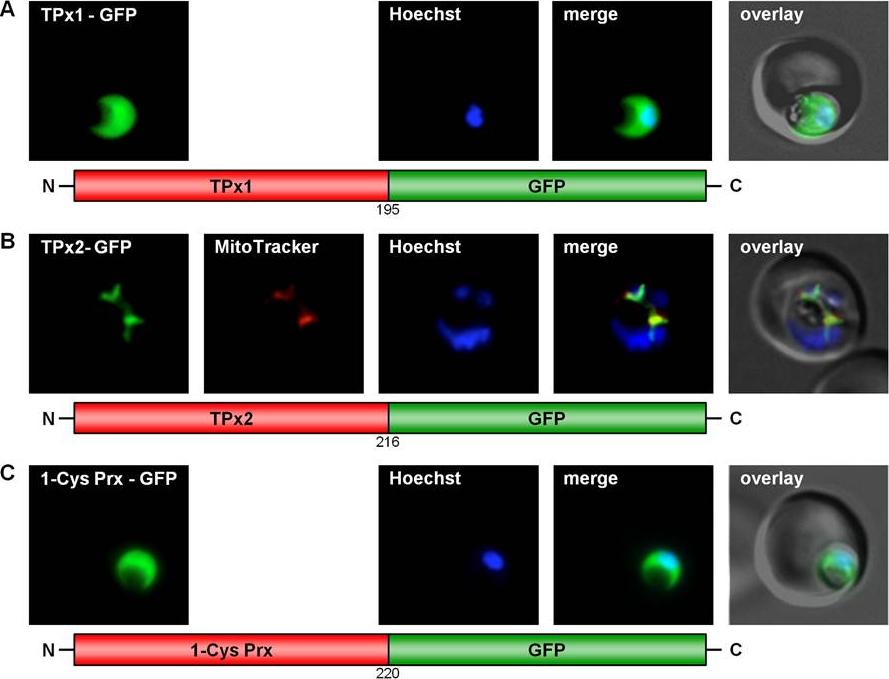
GFP targeting by various P. falciparum peroxiredoxins. (A) Cytosolic localization of TPx1. (B) Mitochondrial targeting of TPx2. (C) Cytosolic localization of 1-Cys Prx. Live cell imaging of erythrocytes infected with transgenic parasites for solely cytosolic GFP signals. Colocalization of GFP with the mitochondrial stain MitoTrackerOrange in fixed cells.Kehr S, Sturm N, Rahlfs S, Przyborski JM, Becker K. Compartmentation of redox metabolism in malaria parasites. PLoS Pathog. 2010 6:e1001242.
See original on MMP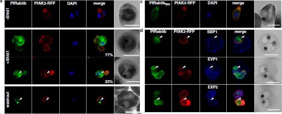
Localization of PfRab5bQ94L-YFP-DD to the TVN. a Time-lapse imaging of PfRab5bQ94L-YFP-DD (green) and TR-ceramide (red) fluorescence after stabilization with Shld1. PfRab5bQ94L-YFP-DD and TR-ceramide was co-localized to a rapidly moving compartment (arrowheads). b The pseudocolors of TR-ceramide at 0, 100, and 200 s were converted to red, green and blue, respectively, and then merged into a single frame. Several extended, mobile TVNs are shown in each color (arrowhead as an example), whereas a stable PVM is shown in white. (c) Cells expressing PfRab5bQ94L-YFP-DD (green) were stained with anti-PfTPx-1 antibody (red) and DAPI (blue). Cytoplasmic PfTPx-1 was not detected from the TVN, where PfRab5bQ94L-YFP-DD (arrowheads) localizes. Bars 5 μmץEbine K, Hirai M, Sakaguchi M, Yahata K, Kaneko O, Saito-Nakano Y. Plasmodium Rab5b is secreted to the cytoplasmic face of the tubovesicular network ininfected red blood cells together with N-acylated adenylate kinase 2. Malar J. 2016 15:323.
See original on MMP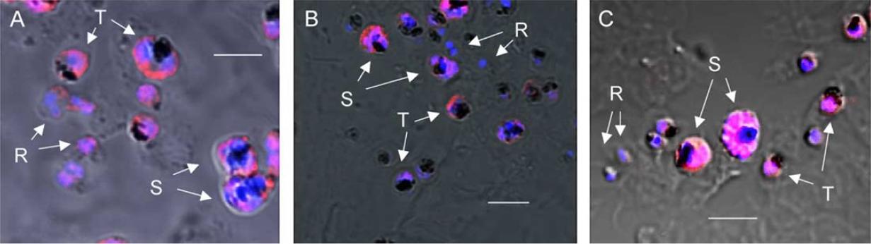
The expression of PfTPx-1 (A), Pf1-Cys-Prx (B), and PfTrx-1 (C) proteins in P. falciparum cells during the erythrocytic stage. Parasite cells in panels (A–C) are stained with antiserum specific for each antioxidant protein (Alexa Fluor 568, red) and TO-PRO-3 iodide (blue; nucleolus staining). Letters with arrows are ring stage (R), schizont (S), and late trophozoite (T). Immunofluorescence microscopy of P. falciparum erythrocytic-stage parasites revealed that PfTPx-1 was expressed in the parasite cytoplasm during the ring, trophozoite and schizont stages. PfTPx-1 thus showed constitutive expression through the erythrocytic stage A). In contrast, significant staining of Pf1-Cys-Prx was detected in the trophozoites in the cytoplasm of the parasite. Expression of Pf1-Cys-Prx in late schizonts was weak in comparison to that of PfTPx-1 in parasites of the same stage, and it was not detected in the ring stage. Pf1-Cys-Prx thus showed limited expression during the trophozoite and early schizont stages (B). Significant expression of PfTrx-1 was observed after the trophozoite stage, but expression was weak during the ring stage (C).Yano K, Komaki-Yasuda K, Kobayashi T, Takemae H, Kita K, Kano S, Kawazu S. Expression of mRNAs and proteins for peroxiredoxins in Plasmodium falciparum erythrocytic stage. Parasitol Int. 2005 Mar;54(1):35-41.
See original on MMPMore information
| PlasmoDB | PF3D7_1438900 |
| GeneDB | PF3D7_1438900 |
| Malaria Metabolic Pathways | Localisation images Pathways mapped to |
| Previous ID(s) | PF14_0368 |
| Orthologs | PBANKA_1302800 , PCHAS_1306000 , PKNH_1243400 , PVP01_1268100 , PVX_118545 , PY17X_1306600 |
| Google Scholar | Search for all mentions of this gene |