PF3D7_1437900 HSP40, subfamily A, putative (ERdj3)
Disruptability [+]
| Species | Disruptability | Reference | Submitter |
|---|---|---|---|
| P. falciparum 3D7 |
Possible |
USF piggyBac screen (Insert. mut.) | USF PiggyBac Screen |
Mutant phenotypes [+]
None reported yet. Please press the '+' button above to add one.Imaging data (from Malaria Metabolic Pathways)
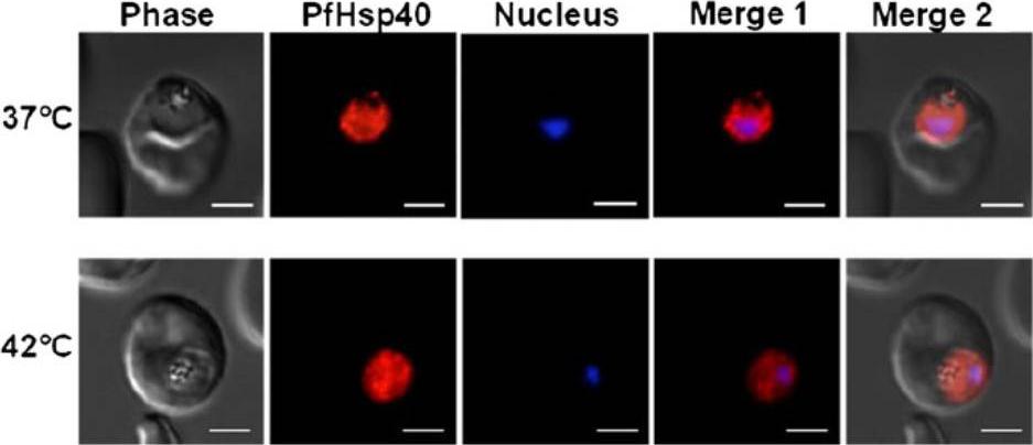
PfHsp40 resides mainly in the parasite cytosol. Immunofluorescence staining to detect PfHsp40 was conducted on trophozoite stage P. falciparum-infected erythrocytes. Upper panels parasite infected erythrocytes maintained at 37°C, lower panels parasite infected erythrocytes incubated at 42°C for 2 h prior to fixation and staining. PfHsp40 was detected using the rabbit anti-PfHsp40 antibody and Cy3-conjugated goat anti-rabbit secondary antibody (indicated in red). Parasite nuclei were stained with Hoechst (indicated in blue). Columns: Phase phase-contrast image, Nucleus parasite nuclei, PfHsp40 PfHsp40 localization, Merge 1 merged image indicating PfHsp40 localization relative to the parasite nucleus. The white size bars in each frame indicate 3 μm.Botha M, Chiang AN, Needham PG, Stephens LL, Hoppe HC, Külzer S, Przyborski JM, Lingelbach K, Wipf P, Brodsky JL, Shonhai A, Blatch GL. Plasmodium falciparum encodes a single cytosolic type I Hsp40 that functionally interacts with Hsp70 and is upregulated by heat shock. Cell Stress Chaperones. 2011 16(4):389-401.
See original on MMP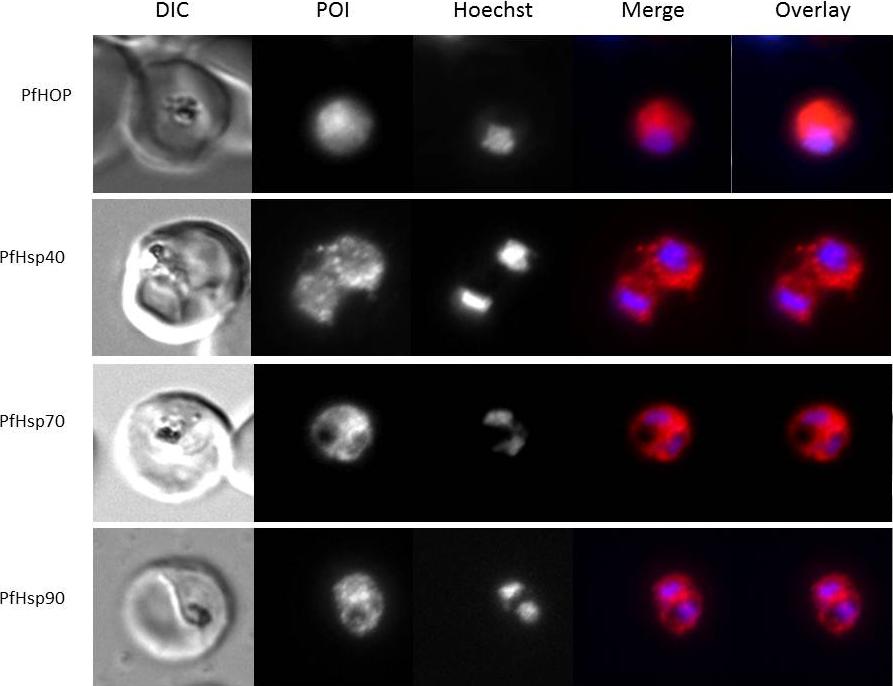
Immunofluorescence staining to detect Hsp40, HSP70, HSP90 and HOP was conducted on trophozoite stage P. falciparum-infected erythrocytes. Panels show from left to right a DIC image, distribution of protein of interest (POI), nuclear stain (Hoechst), merge and overlay (localization relative to the parasite nucleus and phase-contrast image). All proteins showed cytosolic localization.Botha M, Chiang AN, Needham PG, Stephens LL, Hoppe HC, Külzer S, Przyborski JM, Lingelbach K, Wipf P, Brodsky JL, Shonhai A, Blatch GL. Plasmodium falciparum encodes a single cytosolic type I Hsp40 that functionally interacts with Hsp70 and is upregulated by heat shock. Cell Stress Chaperones. 2011 16(4):389-401. Pictures were kindly provided by Jude Przyborski.
See original on MMPMore information
| PlasmoDB | PF3D7_1437900 |
| GeneDB | PF3D7_1437900 |
| Malaria Metabolic Pathways | Localisation images Pathways mapped to |
| Previous ID(s) | PF14_0359 |
| Orthologs | PBANKA_0610900 , PCHAS_0612600 , PKNH_0424600 , PVP01_1311600 , PVX_084650 , PY17X_0613400 |
| Google Scholar | Search for all mentions of this gene |