PF3D7_1436300 translocon component PTEX150 (PTEX150)
Disruptability [+]
| Species | Disruptability | Reference | Submitter | |
|---|---|---|---|---|
| P. falciparum 3D7 |
Refractory |
19536257 | Theo Sanderson, Wellcome Trust Sanger Institute | |
| P. falciparum 3D7 |
Possible |
USF piggyBac screen (Insert. mut.) | USF PiggyBac Screen | |
| P. berghei ANKA |
Refractory |
RMgm-946 | Imported from RMgmDB | |
| P. berghei ANKA |
Refractory |
RMgm-915 | Imported from RMgmDB | |
| P. berghei ANKA |
Refractory |
RMgm-297 | Imported from RMgmDB | |
| P. berghei ANKA |
Refractory |
PlasmoGEM (Barseq) | PlasmoGEM | |
Mutant phenotypes [+]
| Species | Stage | Phenotype | Reference | Submitter |
|---|---|---|---|---|
| P. falciparum 3D7 | Asexual |
Attenuated |
25043043 (Conditional)
Failure to proliferate and reduced protein export upon glucosamine addition to glmS PTEX150. |
Theo Sanderson, Wellcome Trust Sanger Institute |
Imaging data (from Malaria Metabolic Pathways)
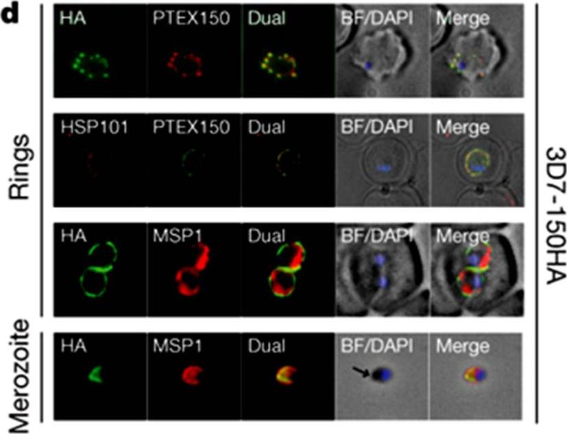
HSP101 and PTEX150 co-localize and have dual apical merozoite and PVM localization. Double labelling IFA on fixed 3D7-150HA ring- and merozoite-stage parasites using the antibodies as indicated. 3D7-150HA is a transgenic P. falciparum (3D7) parasite line where the endogenous gene was modified at its carboxy terminus to include a triple haemagglutinin (HA) epitope tag. The arrows indicate the apical end of the merozoite. BF, bright-field; DAPI, nucleic acid stain 49,6-diamidino-2-phenylindole. PTEX150 and HSP101 co-localize and are found in discrete foci in the membranes surrounding the ring-stage parasite. This membrane is the PVM, as fluorescence surrounds the parasite membrane marker MSP1 (PFI1475w).de Koning-Ward TF, Gilson PR, Boddey JA, Rug M, Smith BJ, Papenfuss AT, Sanders PR, Lundie RJ, Maier AG, Cowman AF, Crabb BS. A newly discovered protein export machine in malaria parasites. Nature. 2009 459(7249):945-9.
See original on MMP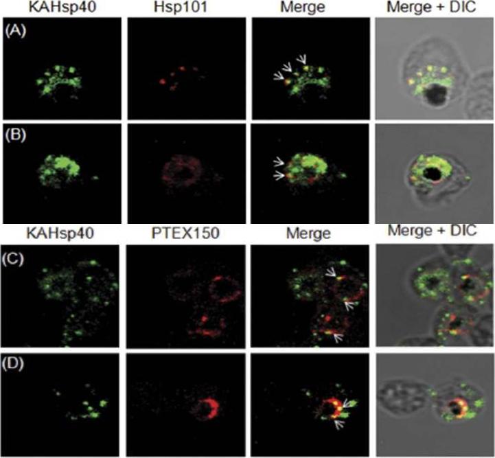
KAHsp40 associates with the PEXEL translocon on the PVM. (A, B) IFA analysis reveals that KAHsp40 and Hsp101 co-localize with each other on the PVM. (C, D) IFA analysis reveals that KAHsp40 and PTEX150 co-localize with each other on the PVM. White arrows indicate the discrete foci in which they co-localize.Acharya P, Chaubey S, Grover M, Tatu U. An Exported Heat Shock Protein 40 Associates with Pathogenesis-Related Knobs in Plasmodium falciparum Infected Erythrocytes. PLoS One. 2012;7(9):e44605.
See original on MMP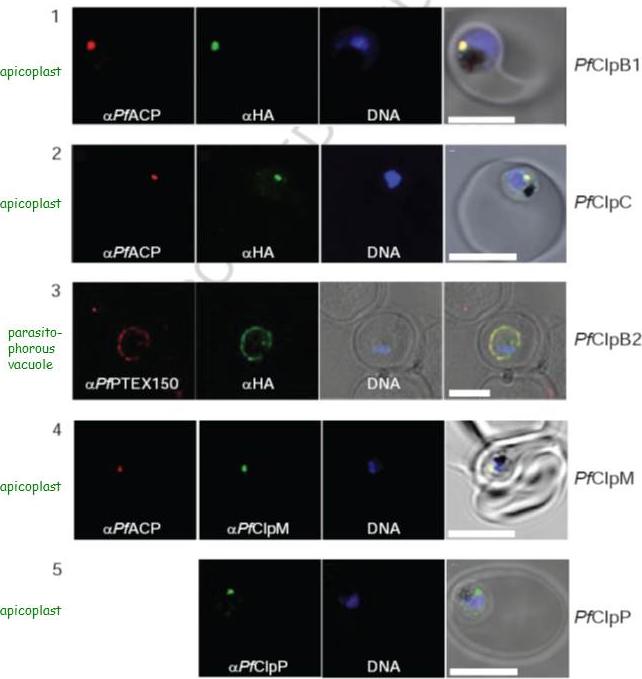
Localization of PfClpB1, PfClpC, PfClpB2, PfClpM, and PfClpP of P. falciparum was tested by immunofluorescent colocalization. In panels 1 and 2, PfClpB1-Strp-3×HA or PfClpC-Strp-3×HA parasites were probed with the apicoplast marker anti-ACP PFB0385w antisera (red), with anti-HA antisera (green), and with Hoescht 33342 to stain for DNA (blue). In panel 3, PfClpB2-Strp-3×HA parasites were probed with the parasitophorous vacuole marker anti-PTEX150 antisera (red), with anti-HA antisera (green), and with Hoescht 33342 (blue). In panels 4 and 5, wild type 3D7 strain was probed with anti-ACP antisera (red), with anti-PfClpM antisera or anti-ClpP antisera (green), and with Hoescht 33342 (blue). For all panels, the right most image represents the merged fluorescence images with the DIC or transmission image. The scale bars correspond to 5 mm.El Bakkouri M, Pow A, Mulichak A, Cheung KL, Artz JD, Amani M, Fell S, de Koning T, Goodman CD, McFadden GI, Ortega J, Hui R, Houry WA. The Clp chaperones and proteases of the human malaria parasite Plasmodium falciparum. J Mol Biol. 2010 404(3):456-77
See original on MMP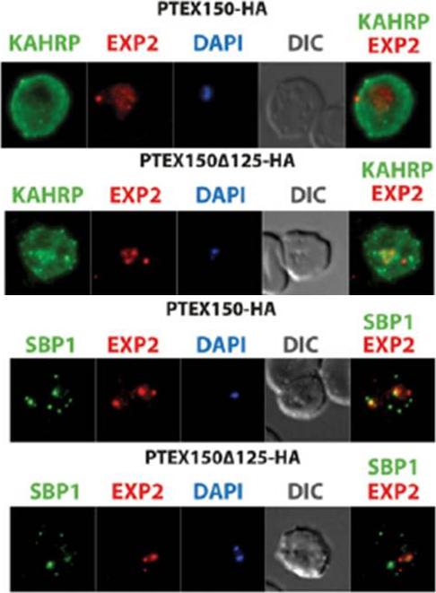
The export of KAHRP and SBP1 in the PTEX150Δ125-HA mutant compared to full length PTEX150-HA parasites. PTEX150-HA and PTEX150Δ125-HA parasites were synchronized to an invasion window of 5 h. The regions occupied by the parasite are indicated by DAPI and EXP2 staining. KAHRP is a PEXEL protein, which initially displays a diffuse staining of the erythrocyte cytosol before accumulating in punctate regions below the plasma membrane of the host cell. SBP is a PNEP that localizes to punctate vesicular structures called Maurer’s clefts (MC) in the host cytosol. IFAs were counterstained with antibodies to EXP2 to delineate where the boundaries of the parasite. No significant difference between the PTEX150Δ125-HA and PTEX150-HA parasite lines was observed.Elsworth B, Sanders PR, Nebl T, Batinovic S, Kalanon M, Nie CQ, Charnaud SC, Bullen HE, de Koning Ward TF, Tilley L, Crabb BS, Gilson PR. Proteomic analysis reveals novel proteins associated with the Plasmodium protein exporter PTEX and a loss of complex stability upon truncation of the core PTEX component, PTEX150. Cell Microbiol. 2016 18(11):1551-1569.
See original on MMPMore information
| PlasmoDB | PF3D7_1436300 |
| GeneDB | PF3D7_1436300 |
| Malaria Metabolic Pathways | Localisation images Pathways mapped to |
| Previous ID(s) | PF14_0344, Pf112 |
| Orthologs | PBANKA_1008500 , PCHAS_1009400 , PKNH_0422900 , PVP01_1313200 , PVX_084720 , PY17X_1010100 |
| Google Scholar | Search for all mentions of this gene |