PF3D7_1422500 ERAD-associated E3 ubiquitin-protein ligase HRD1 (HRD1)
Disruptability [+]
| Species | Disruptability | Reference | Submitter |
|---|---|---|---|
| P. falciparum 3D7 |
Refractory |
22912882 | Theo Sanderson, Wellcome Trust Sanger Institute |
| P. falciparum 3D7 |
Refractory |
USF piggyBac screen (Insert. mut.) | USF PiggyBac Screen |
Mutant phenotypes [+]
None reported yet. Please press the '+' button above to add one.Imaging data (from Malaria Metabolic Pathways)
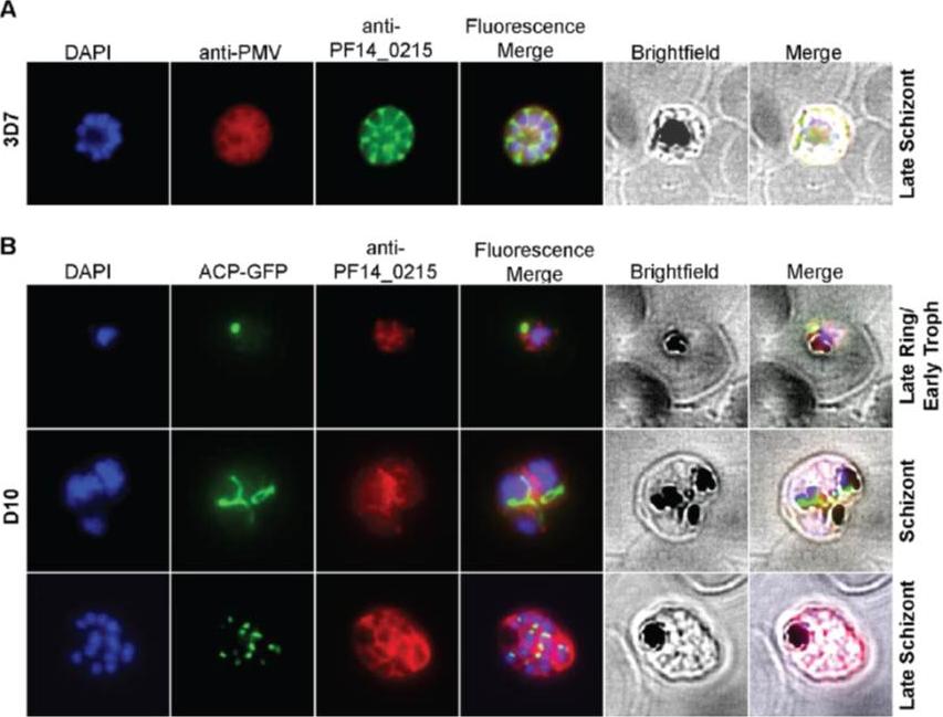
Cellular localization of HRD1. (A) HRD1 and plasmepsin V (PMV), an ER membrane marker, co-localize in P. falciparum. (B) At various stages of the parasite life cycle, HRD1 was co-stained with the nuclei (DAPI) and the apicoplast (ACP-GFP) in the P. falciparum strain D10. HRD1 was found within reticular structures outside the nuclear regions at the trophozoite and schizont stages of the parasite, similar to the physical attributes of the ER. In the late schizont stage HRD1 proteins reside within globular structures surrounding each budding merozoite in a pattern typical of the ER. These observations support that HRD1-E3 ubiquitin ligase resides in the ER membrane, consistent with a putative role in the ERAD pathway.Chung DW, Ponts N, Prudhomme J, Rodrigues EM, Le Roch KG. Characterization of the Ubiquitylating Components of the Human Malaria Parasite's Protein Degradation Pathway. PLoS One. 2012;7(8):e43477.
See original on MMPMore information
| PlasmoDB | PF3D7_1422500 |
| GeneDB | PF3D7_1422500 |
| Malaria Metabolic Pathways | Localisation images Pathways mapped to |
| Previous ID(s) | PF14_0215 |
| Orthologs | PBANKA_1020900 , PCHAS_1021700 , PKNH_1335500 , PVP01_1326500 , PVX_085355 , PY17X_1022800 |
| Google Scholar | Search for all mentions of this gene |