PF3D7_1420700 surface protein P113 (P113)
Disruptability [+]
| Species | Disruptability | Reference | Submitter | |
|---|---|---|---|---|
| P. falciparum 3D7 |
Refractory |
22986493 | Theo Sanderson, Wellcome Trust Sanger Institute | |
| P. falciparum 3D7 |
Possible |
USF piggyBac screen (Insert. mut.) | USF PiggyBac Screen | |
| P. berghei ANKA |
Possible |
RMgm-1001 | Imported from RMgmDB | |
Mutant phenotypes [+]
| Species | Stage | Phenotype | Reference | Submitter |
|---|---|---|---|---|
| P. falciparum 3D7 | Asexual |
Possible |
35403274 (Knock down)
\"Genetic knockdown or partial deletion of P113 did not significantly reduce parasite growth or protein export but did disrupt the morphology of the PVM, suggesting that P113 may play a role in maintaining normal PVM architecture.\" |
Theo Sanderson, Francis Crick Institute |
| P. falciparum 3D7 | Asexual |
Possible |
35403274 \"Genetic knockdown or partial deletion of P113 did not significantly reduce parasite growth or protein export but did disrupt the morphology of the PVM, suggesting that P113 may play a role in maintaining normal PVM architecture.\" |
Theo Sanderson, Francis Crick Institute |
| P. berghei ANKA | Asexual |
No difference |
RMgm-1001 | Imported from RMgmDB |
| P. berghei ANKA | Oocyst |
No difference |
RMgm-1001 | Imported from RMgmDB |
| P. berghei ANKA | Sporozoite |
No difference |
RMgm-1001 | Imported from RMgmDB |
| P. berghei ANKA | Liver |
Difference from wild-type |
RMgm-1001
Sporozoites show normal gliding motility, attachment to hepatocytes and invasion of hepatocytes (possibly an enhanced invasion rate). Reduced liver stage development as shown by a prolonged pre-patent period (possibly due to reduced transformation of intracellular sporozoites into early liver stages and reduced production of merozoites). |
Imported from RMgmDB |
Imaging data (from Malaria Metabolic Pathways)
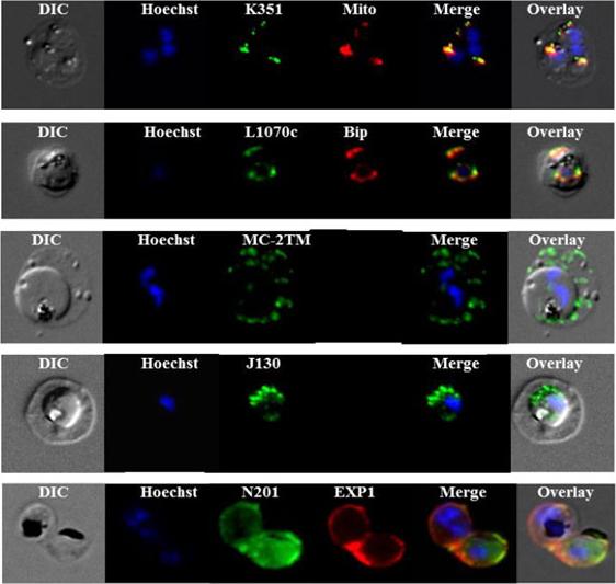
Sub-cellular localization of P. falciparum DRM-associated proteins. Immunofluorescence studies were performed on paraformaldehyde-fixed P.falciparum trophozoite-iRBCs using mouse immune sera raised against the DRM-associated proteins PFJ130, (PF10_0130) PFN201, (PF14_0201 surface protein) PFB985c, (PFB0985c) Pfmc-2TM Maurer's cleft two transmembrane protein; PFL1070c, endoplasmin homolog precursor) PFK351 (PF11_0351) HSP70. Immune sera specific for PfBip and PfEXP1-were used for co-labeling, as indicated. Mitochondria labeling was performed with MitoTracker® probe. Parasite nuclei were stained with Hoechst 33342. Microscopic observations were made on an apotome-equipped Axio Imager 2 (Carl Ziess, Jena) with a 100× apochromat objective and DIC. The differential interference contrast (DIC), merged and overlay images are presented. Bar size: 5 µm. PfK351overlays mitotracker thus localized to parasite mitochondrion. PfL1070 colocalizes with the ER-resident marker PfBiP. PfK55, a conserved hypothetical protein with unknown function, contains a SP and a ER retention signal (KDEL), but due to poor IFA, the exact subcellular localization of this protein remained elusive.Yam XY, Birago C, Fratini F, Di Girolamo F, Raggi C, Sargiacomo M, Bachi A, Berry L, Fall G, Curra C, Pizzi E, Braun-Breton C, Ponzi M. Proteomic analysis of detergent-resistant membrane microdomains in trophozoite blood stage of the human malaria parasite Plasmodium falciparum. Mol Cell Proteomics. 2013 12(12):3948-61
See original on MMP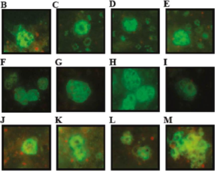
Fixed and blocked late schizonts and ring stages were incubated with rabbit anti-Pf92 immune sera (B-I) or with Pf113 immune rabbit sera (J-M). In late schizonts Pf92 localized in merozoite and in ring stages (C-E). Pf113 was only detected on the merozoite surface, and not in other subcellular compartments or in any other intraerythrocytic lifecycle stage.Obando-Martinez AZ, Curtidor H, Arévalo-Pinzón G, Vanegas M, Vizcaino C, Patarroyo MA, Patarroyo ME. Conserved high activity binding peptides are involved in adhesion of two detergent-resistant membrane-associated merozoite proteins to red blood cells during invasion. J Med Chem. 2010 53:3907-3918.
See original on MMP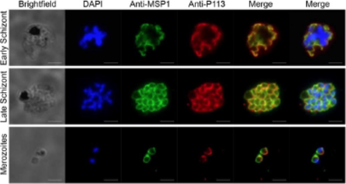
P113 is expressed in both early and late-stage P. falciparum schizonts and on the merozoite surface. Fixed blood stage P. falciparum schizonts and free merozoites were co-stained with antibodies raised to P113 and the merozoite surface marker anti-MSP1 (a), or an inner membrane complex marker, MTIP (b) as indicated. Nucleic acid is stained with DAPI and scale bars represent 3μm.Galaway F, Drought LG, Fala M, Cross N, Kemp AC, Rayner JC, Wright GJ. P113 is a merozoite surface protein that binds the N terminus of Plasmodium falciparum RH5. Nat Commun. 2017 8:14333.
See original on MMP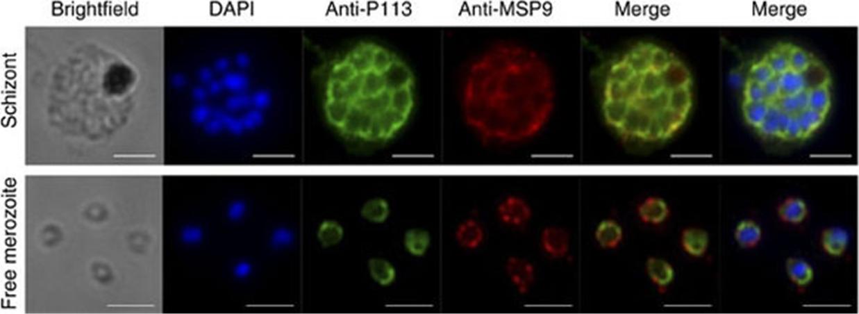
P113 is expressed in schizonts and on the surface of free merozoites. Fixed blood-stage P. falciparum schizonts and free merozoites were probed with anti-P113 antibodies (green), the merozoite surface marker anti-MSP9 (red) and nucleic acid stained with DAPI (blue). Scale bars, 3 mm. P113, is expressed in early and late-stage schizonts and on the surface of free merozoites by co-staining with the established merozoite surface markers MSP9.Galaway F, Drought LG, Fala M, Cross N, Kemp AC, Rayner JC, Wright GJ. P113 is a merozoite surface protein that binds the N terminus of Plasmodium falciparum RH5. Nat Commun. 2017 8:14333. PMID: 28186186
See original on MMPMore information
| PlasmoDB | PF3D7_1420700 |
| GeneDB | PF3D7_1420700 |
| Malaria Metabolic Pathways | Localisation images Pathways mapped to |
| Previous ID(s) | PF14_0201, Pf113 |
| Orthologs | PBANKA_1022500 , PCHAS_1023300 , PKNH_1337300 , PVP01_1328200 , PVX_085445 , PY17X_1024400 |
| Google Scholar | Search for all mentions of this gene |