PF3D7_1417800 DNA replication licensing factor MCM2 (MCM2)
Disruptability [+]
| Species | Disruptability | Reference | Submitter | |
|---|---|---|---|---|
| P. falciparum 3D7 |
Refractory |
USF piggyBac screen (Insert. mut.) | USF PiggyBac Screen | |
| P. berghei ANKA |
Refractory |
PlasmoGEM (Barseq) | PlasmoGEM | |
Mutant phenotypes [+]
None reported yet. Please press the '+' button above to add one.Imaging data (from Malaria Metabolic Pathways)
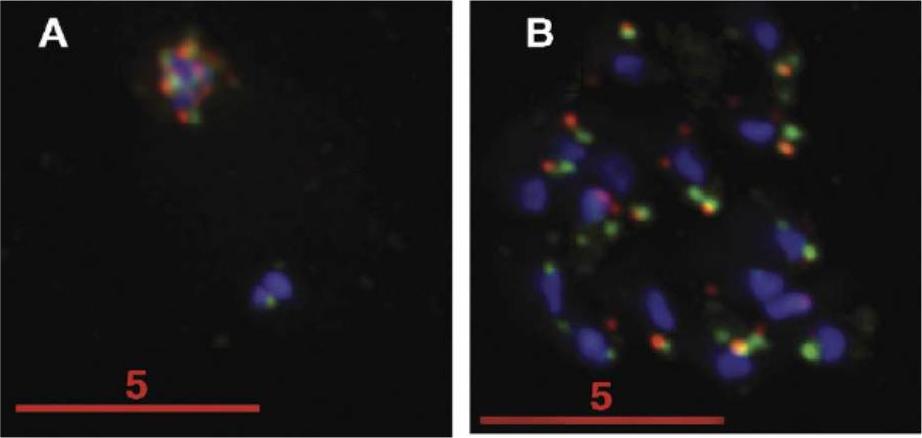
Parasites on coverslips were fixed with paraformaldehyde and permeabilized with 0.05% saponin. The cells were probed with PfMCM2 and PfMCM6 antibodies followed by Alexafluor 555 labeled antimouse (green, PfMCM2) and Alexafluor 647 labeled anti-rabbit (red, PfMCM6) secondary antibodies. DNA was stained with DAPI (blue). Images were analyzed by DeltaVision deconvolution microscopy. The bar indicates 5mM. PfMCM2, 6, and 7 appear localized in both cytosolic and nucleosolic fractions during all intraerythrocytic stages of P. falciparum development, with increased nuclear localization in schizonts. Patterson S, Robert C, Whittle C, Chakrabarti R, Doerig C, Chakrabarti D. Pre-replication complex organization in the atypical DNA replication cycle of Plasmodium falciparum: characterization of the mini-chromosome maintenance (MCM) complex formation. Mol Biochem Parasitol. 2006 145:50-9. Copyright Elsevier 2009.
See original on MMP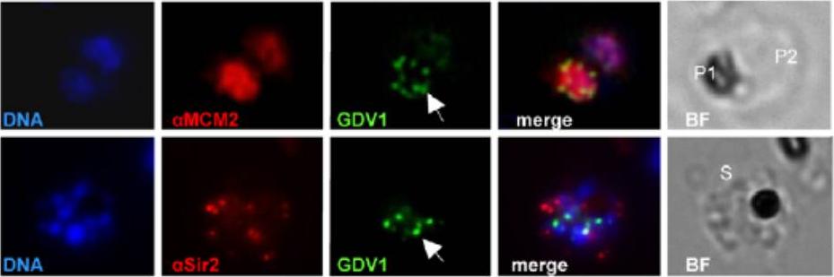
Subcellular localization of PfGDV1 (this gene product is expressed in gametocytes and is not included in MPMP). Parasites transformed with GFP- PfGDV1 were stained with DAPI (DNA stain) and the indicated anti-sera, and then examined by fluorescence microscopy (Zeiss Axiovert 200, 1000xmagnification). Images are shown of the DAPI stain (DNA), GFP-tagged PfGDV1 epifluorescence (GDV1), and antibodies specific for the indicated proteins. The corresponding merged and bright field (BF) images are included on the right. PfGDV1 expression is indicated with an arrow; locations of parasites in the BF image are indicated with a P for parasite or S for schizont. Colocalization of PfGDV1 with nuclear proteins. A doubly infected erythrocyte (Upper) with one parasite in the plane of the image (P1) and the other below (P2). Both P1 and P2 are positive for GDV1 and aMCM2. A schizont (S) expressing GDV1 stained with aSir2. MCM2 localized to the nucleus, and PfSir2 to the nuclear periphery.Eksi S, Morahan BJ, Haile Y, Furuya T, Jiang H, Ali O, Xu H, Kiattibutr K, Suri A, Czesny B, Adeyemo A, Myers TG, Sattabongkot J, Su XZ, Williamson KC. Plasmodium falciparum gametocyte development 1 (Pfgdv1) and gametocytogenesis early gene identification and commitment to sexual development. PLoS Pathog. 2012 8(10):e1002964.
See original on MMP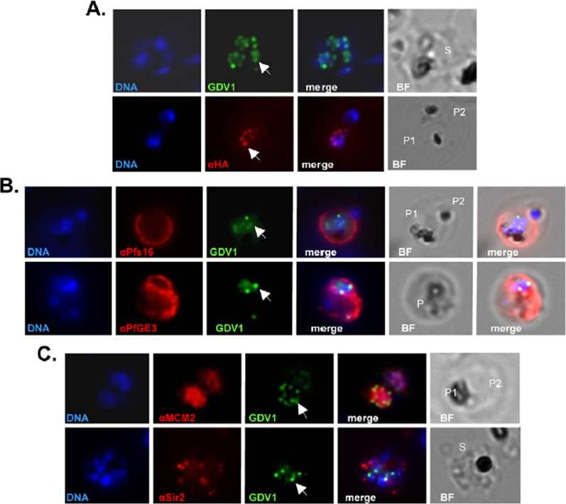
Subcellular localization of PfGDV1. Parasites transformed with GFP- or HA-tagged PfGDV1 were stained with DAPI (DNA stain) and the indicated anti-sera, and then examined by fluorescence microscopy. Images are shown of the DAPI stain (DNA), GFP-tagged PfGDV1 epifluorescence (GDV1), and antibodies specific for HA (aHA), Pfs16 (aPfs16), PfGE3 (aPfGE3), PfMCM2 (aMCM2), and PfSir2 (aSir2). The corresponding merged and bright field (BF) images are included on the right. PfGDV1 expression is indicated with an arrow; locations of parasites in the BF image are indicated with a P for parasite or S for schizont. A) A schizont (S) (Upper) expressing GDV1 and a doubly infected erythrocyte (Lower) with one parasite (P1) expressing HA-tagged PfGDV1 (aHA) and another negative (P2) for anti-HA antibodies. B) Costaining of parasites expressing GDV1 with early gameto-cytogenesis markers. A doubly infected erythrocyte (Upper) with one parasite (P1) positive for GDV1 and aPfs16 and the other (P2) negative for both. An erythrocyte (Lower) infected with a parasite (P) positive for GDV1 and aPfGE3. C) Colocalization of PfGDV1 with nuclear proteins. A doubly infected erythrocyte (Upper) with one parasite in the plane of the image (P1) and the other below (P2). Both P1 and P2 are positive for GDV1 and aMCM2. A schizont (S) (Lower) expressing GDV1 stained with aSir2.Eksi S, Morahan BJ, Haile Y, Furuya T, Jiang H, Ali O, Xu H, Kiattibutr K, Suri A, Czesny B, Adeyemo A, Myers TG, Sattabongkot J, Su XZ, Williamson KC. Plasmodium falciparum gametocyte development 1 (Pfgdv1) and gametocytogenesis early gene identification and commitment to sexual development. PLoS Pathog. 2012;8(10):e1002964.
See original on MMPMore information
| PlasmoDB | PF3D7_1417800 |
| GeneDB | PF3D7_1417800 |
| Malaria Metabolic Pathways | Localisation images Pathways mapped to |
| Previous ID(s) | PF14_0177 |
| Orthologs | PBANKA_1024900 , PCHAS_1025700 , PKNH_1340200 , PVP01_1331100 , PVX_085565 , PY17X_1027300 |
| Google Scholar | Search for all mentions of this gene |