PF3D7_1408000 plasmepsin II
Disruptability [+]
| Species | Disruptability | Reference | Submitter | |
|---|---|---|---|---|
| P. falciparum 3D7 |
Possible |
15491999 aberrant moitochondrial morphology |
Theo Sanderson, Wellcome Trust Sanger Institute | |
| P. falciparum 3D7 |
Refractory |
USF piggyBac screen (Insert. mut.) | USF PiggyBac Screen | |
| P. berghei ANKA |
Possible |
RMgm-808 | Imported from RMgmDB | |
| P. berghei ANKA |
Possible |
RMgm-314 | Imported from RMgmDB | |
| P. berghei ANKA |
Possible |
RMgm-315 | Imported from RMgmDB | |
| P. berghei ANKA |
Possible |
RMgm-316 | Imported from RMgmDB | |
| P. berghei ANKA |
Possible |
PlasmoGEM (Barseq) | PlasmoGEM | |
Mutant phenotypes [+]
| Species | Stage | Phenotype | Reference | Submitter |
|---|---|---|---|---|
| P. berghei ANKA | Asexual |
Difference from wild-type |
RMgm-808
Asexual blood stages show a (slightly) reduced growth rate and reduced hemozoin formation. |
Imported from RMgmDB |
| P. berghei ANKA | Asexual |
Difference from wild-type |
RMgm-316
Asexual blood stages show a slightly reduced growth rate and show a virulence-attenuated phenotype in mice (see 'Additional remarks phenotype'). |
Imported from RMgmDB |
| P. berghei ANKA | Asexual |
Difference from wild-type |
RMgm-314
Asexual blood stages show a slightly reduced growth rate and show a virulence-attenuated phenotype in mice (see 'Additional remarks phenotype'). |
Imported from RMgmDB |
| P. berghei ANKA | Asexual |
Difference from wild-type |
RMgm-315
Asexual blood stages show a slightly reduced growth rate and show a virulence-attenuated phenotype in mice (see 'Additional remarks phenotype'). |
Imported from RMgmDB |
| P. berghei ANKA | Asexual |
Attenuated |
PlasmoGEM (Barseq) | PlasmoGEM |
| P. berghei ANKA | Gametocyte |
No difference |
RMgm-316 | Imported from RMgmDB |
| P. berghei ANKA | Ookinete |
No difference |
RMgm-316 | Imported from RMgmDB |
| P. berghei ANKA | Oocyst |
No difference |
RMgm-316 | Imported from RMgmDB |
| P. berghei ANKA | Sporozoite |
No difference |
RMgm-316 | Imported from RMgmDB |
| P. berghei ANKA | Liver |
No difference |
RMgm-316 | Imported from RMgmDB |
Imaging data (from Malaria Metabolic Pathways)
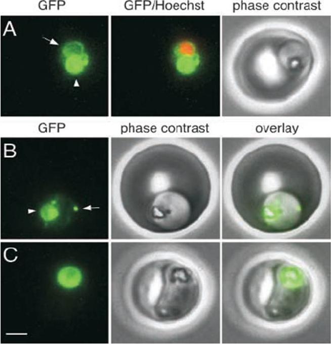
A trophozoite exhibiting GFP fluorescence in the food vacuole (arrowhead) and in a perinuclear ring (arrow). Fluorescence from the nuclear stain Hoechst 33342 is pseudocolored red. (B) A trophozoite with a fluorescent food vacuole (arrowhead) and a bright fluorescent spot (arrow) that lies at the periphery of the parasite. (C) A mature parasite displaying a large fluorescent food vacuole. Note the absence of fluorescence outside of the food vacuole. Bar, 2 mm.Klemba et al., 2004. Originally published in The Journal of Cell Biology. doi:10.1083/jcb200307147. Klemba M, Beatty W, Gluzman I, Goldberg DE. Trafficking of plasmepsin II to the food vacuole of the malaria parasite Plasmodium falciparum. J Cell Biol. 2004 Jan 5;164(1):47-56.
See original on MMP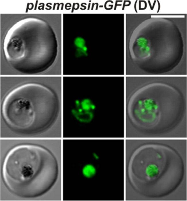
Confocal fluorescence microscopy images of transfected 3D7 P. falciparum-infected RBCs expressing a GFP chimera directed to the digestive vacuole. DIC image, the GFP fluorescence signal, and an overlay of a P. falciparum plasmepsin II-GFP transfectant. Scale bar = 5 µm. En route to the digestive vacuole during the ring and trophozoite stages plasmepsin-GFP is observed in the endoplasmic reticulum and in cytosome-derived vesicles (top and middle rows). In the late stages of parasite development, the chimera accumulates in the digestive vacuole as shown in the bottom row. Tilley L, McFadden G, Cowman A, Klonis N. Illuminating Plasmodium falciparum-infected red blood cells. Trends Parasitol. 2007 23:268-77. PMID: 17434344
See original on MMP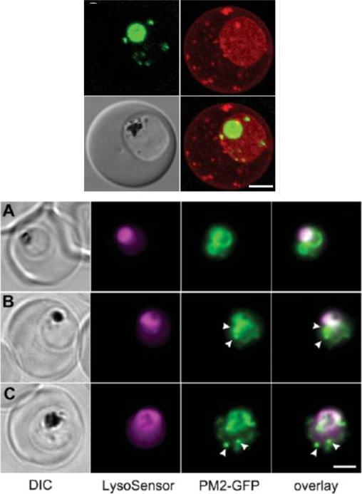
Upper panel: A 3D7-PM2 trophozoite was labelled with BODIPY® ceramide and the GFP (green fluorescence) and BODIPY® (red fluorescence) signals were imaged by confocal microscopy. The images shown represent an average projection obtained from a series of optical sections. BODIPY® ceramide labels the membrane rich parasite cytoplasm as well as punctate structures in the RBC cytosol that have previously been shown to be Maurer’s clefts and other membranous compartments. The cytostomal vesicles are visible as apparently independent structures in the parasite cytoplasm. Lower panel: Mixed-stage transfected parasites were incubated with the pH probe, LysoSensor Blue. Shown are bright field, LysoSensor Blue, GFP and merged GFP/LysoSensor Blue images. The LysoSensor Blue fluorescence is largely associated with the DV, while the GFP chimaera is also present in cytostomal vesicles (arrows) that do not stain with the LysoSensor Blue. Scale bar, 2 μm.Klonis N, Tan O, Jackson K, Goldberg D, Klemba M, Tilley L. Evaluation of pH during cytostomal endocytosis and vacuolar catabolism of haemoglobin in Plasmodium falciparum. Biochem J. 2007 407(3):343-54.
See original on MMP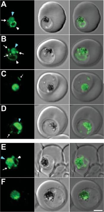
(A–D) 3D7 and (E, F) Dd2 transfectants expressing PM2–GFP were imaged at the young to mid trophozoite (A, B, E) or mature trophozoite to schizont (C, D, F) stage. The images (left to right) represent a GFP fluorescence image, a DIC (differential interference contrast) image and an overlay of these images. DV-located GFP is indicated with a blue arrowhead. ER-located GFP is indicated with white arrowheads, while cytostomal vesicles are indicated with white arrows. Scale bar, 2 mmKlonis N, Tan O, Jackson K, Goldberg D, Klemba M, Tilley L. Evaluation of pH during cytostomal endocytosis and vacuolar catabolism of haemoglobin in Plasmodium falciparum. Biochem J. 2007 407(3):343-54.
See original on MMP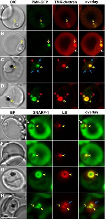
Identification of haemoglobinase-containing and LysoSensor-Blue labelled compartments in infected RBCs. The panels are DIC or bright-field images, GFP or SNARF-1 fluorescence (depicted in green) or TMR-dextran or LysoSensor Blue fluorescence (depicted in red), and overlays of the red and green images. (A-D) Plasmepsin-II-GFP-3D7 transfectants in TMR-dextran containing resealed RBCs. The pre-digestive vacuole (DV) and DV compartments (yellow arrows) and the extra-DV compartments (red arrowheads) contain both plasmepsin-II-GFP and TMR-dextran. Plasmepsin-II-GFP is also present in structures that could represent nascent cytostomal invaginations (blue arrows). There is no plasmepsin-II-GFP associated with a larger spherical profile structure (white arrow). (E-H) In rings (E,F) and trophozoites (G,H) in SNARF-1-dextran containing RBCs, LysoSensor Blue is associated with peripheral structures and the DV (arrowheads) and extra-DV structures (yellow arrows). The spherical structure in ring-stage parasites (A, white arrow) is not labeled. Some additional structures in the trophozoite-stage parasites are weakly labeled with LysoSensor Blue, but do not contain SNARF-1-dextran (H, blue arrow). Scale bars: 5 mm. Abu Bakar N, Klonis N, Hanssen E, Chan C, Tilley L. Digestive-vacuole genesis and endocytic processes in the early intraerythrocytic stages of Plasmodium falciparum. J Cell Sci. 2010 123:441-450.
See original on MMP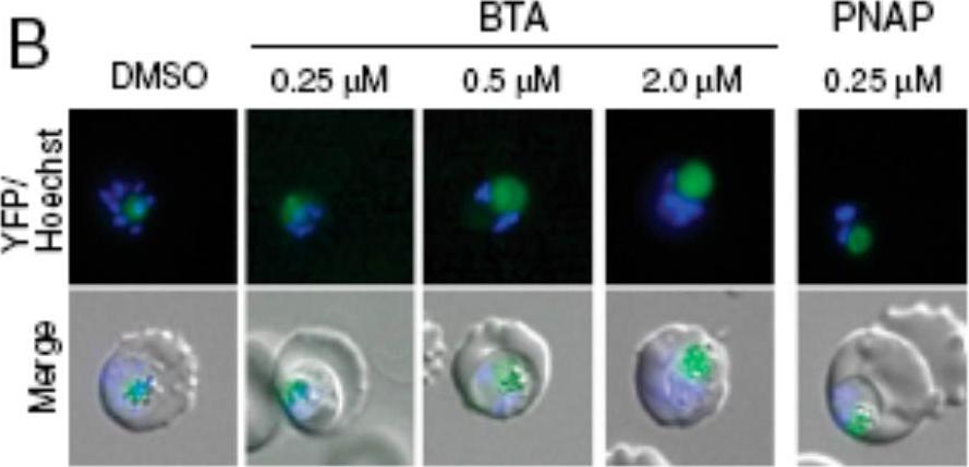
Digestive vacuole (DV) swelling was confirmed by the dose-dependent enlargement of the average DV area. Parasites expressing YFP-tagged plasmepsin II (PMII-YFP), which localizes to the DV, were treated with increasing concentrations of (Benzyl)Tyr-Ala (BTA) and 250 nM Phe-Naphthyl (PNAP) and imaged by fluorescence microscopy. Harbut MB, Velmourougane G, Dalal S, Reiss G, Whisstock JC, Onder O, Brisson D, McGowan S, Klemba M, Greenbaum DC. Bestatin-based chemical biology strategy reveals distinct roles for malaria M1- and M17-family aminopeptidases. Proc Natl Acad Sci U S A. 2011 108:E526-34.
See original on MMP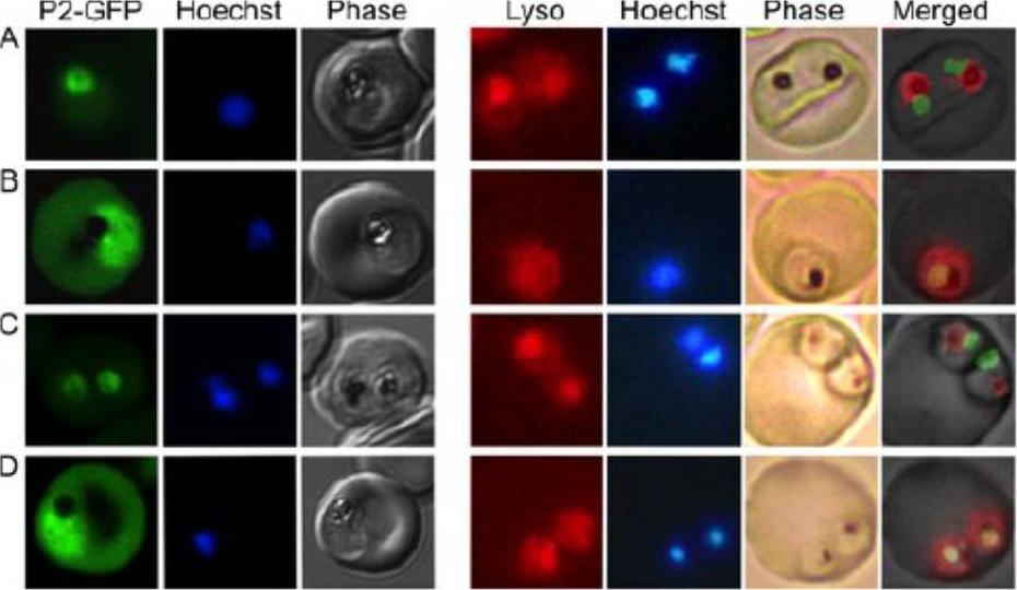
Loss of food vacuolar integrity upon fosmidomycin and wortmannin treatment Confocal fluorescence microscopic localization of a plasmepsin-II-GFP (PMII736 GFP) construct (left panels) or live fluorescence imaging of Lysotracker Red737 stained (right panels) malaria parasites. Control parasites (A) are compared to parasites treated for 24h with either fosmidomycin (+ FSM), fosmidomycin plus geranylgeraniol (+ FSM + GG-ol), or wortmannin (+ wort). Images are representative of at least three independent biological experiments. PMII-GFP was localized to the FV, which is distinguished microscopically in phase images by the presence of dark hemozoin pigment. Following fosmidomycin treatment, PMII-GFP was no longer discretely localized within the FV. Instead, PMII-GFP was dispersed throughout the parasite cytoplasm, consistent with the physical disruption of the FV that was visualized by electron microscopy.Howe R, Kelly M, Jimah J, Hodge D, Odom AR. Isoprenoid biosynthesis inhibition disrupts Rab5 localization and food vacuolar integrity in Plasmodium falciparum. Eukaryot Cell. 2012 12(2):215-23.
See original on MMP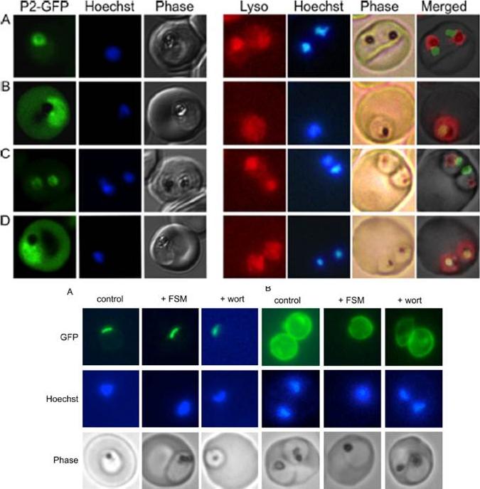
Loss of food vacuolar integrity upon fosmidomycin and wortmannin treatment. Shown is the confocal fluorescence microscopic localization of a plasmepsin II-GFP (P2-GFP) construct (left) or live fluorescence imaging of Lysotracker Red-stained malaria parasites (right). Control parasites (A) are compared to parasites treated for 24 h with either fosmidomycin (B), fosmidomycin plus geranylgeraniol (C), or wortmannin (D). Images are representative of at least three independent biological experiments. Visualization of PMIIGFP in FSM- and wortmannin-treated parasites required higher detector gain levels, resulting in increased observed erythrocyte autofluorescence. Lower panel: Apicoplast and parasitophorous vacuolar targeting in fosmidomycin- and wortmannin-treated parasites. Shown is live-cell fluorescence of malaria parasites that express either the leader sequence (ACPL-GFP) (A) or signal sequence (ACPs-GFP) (B) from P. falciparum acyl carrier protein, fused to GFP, which traffic to the apicoplast or parasitophorous vacuole, respectively. Untreated parasites (control) are compared to fosmidomycin (FSM)- and wortmannin (wort)-treated parasites. Images are representative of at least three independent biological experiments.Howe R, Kelly M, Jimah J, Hodge D, Odom AR. Isoprenoid biosynthesis inhibition disrupts Rab5 localization and food vacuolar integrity in Plasmodium falciparum. Eukaryot Cell. 2013 Feb;12(2):215-23.
See original on MMP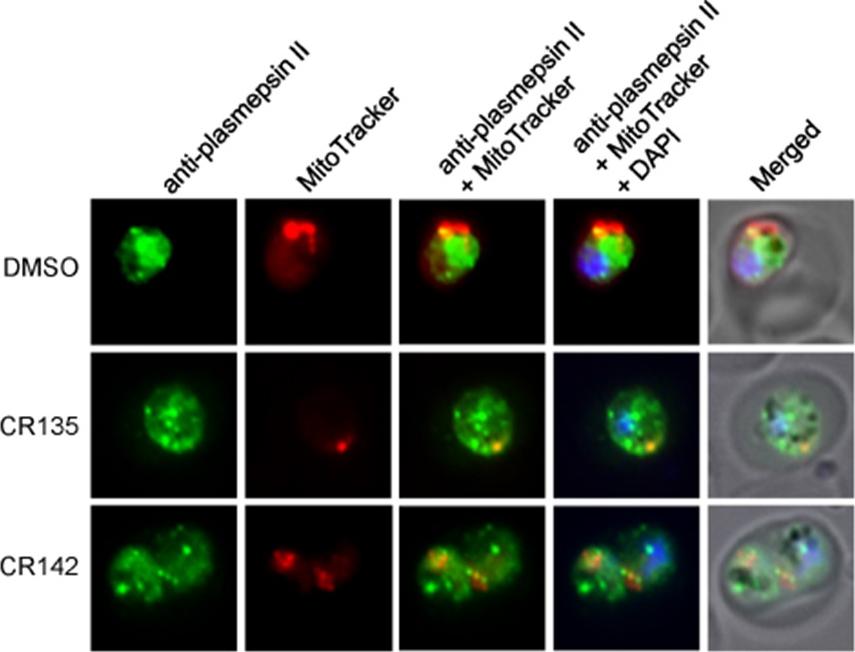
Representative IFA images show food vacuole morphology in parasites treated with vehicle, CR135, and CR142, individually. Anti-Plasmepsin II polyclonal antibody (rabbit) was diluted 1:1000 and an AlexaFluor 488 conjugated anti-rabbit secondary antibody was diluted 1:350. MitoTracker Red stains active mitochondria. Under drug treatment (CR135 or CR142), the globular structure of the food vacuole of a normal parasite was absent, and the vacuole marker Plasmepsin II distributed among many punctate particles scattered throughout the parasite cytosol.Ke H, Morrisey JM, Qu S, Chantarasriwong O, Mather MW, Theodorakis EA, Vaidya AB. Caged Garcinia xanthones: a novel chemical scaffold with potent antimalarial activity. Antimicrob Agents Chemother. 2016 Oct 31. pii: AAC.01220-16PMID:
See original on MMPMore information
| PlasmoDB | PF3D7_1408000 |
| GeneDB | PF3D7_1408000 |
| Malaria Metabolic Pathways | Localisation images Pathways mapped to |
| Previous ID(s) | PF14_0077 |
| Orthologs | PBANKA_1034400 , PCHAS_1035200 , PKNH_1350300 , PVP01_1340900 , PVX_086040 , PY17X_1036800 |
| Google Scholar | Search for all mentions of this gene |