PF3D7_1406700 vacuolar protein sorting-associated protein 29 (VPS29)
Disruptability [+]
| Species | Disruptability | Reference | Submitter | |
|---|---|---|---|---|
| P. falciparum 3D7 |
Refractory |
USF piggyBac screen (Insert. mut.) | USF PiggyBac Screen | |
| P. berghei ANKA |
Refractory |
PlasmoGEM (Barseq) | PlasmoGEM | |
Mutant phenotypes [+]
None reported yet. Please press the '+' button above to add one.Imaging data (from Malaria Metabolic Pathways)
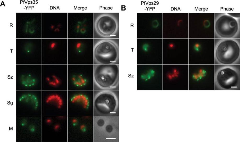
(A) Wide-field epifluorescence images of live parasites expressing PfVps35-YFP. Parasites are shown at ring (R), trophozoite (T), schizont (Sz), segmenter (Sg), and extracellular merozoite (M) stages. Hoechst 33342 fluorescence (DNA) is pseudocolored red. (B) Images of live parasites expressing PfVps29-YFP. Both PfVps29-YFP and PfVps35-YFP were expressed throughout the asexual blood stage. In live parasites expressing PfVps35-YFP, punctate structures were observed in trophozoites and schizonts amid a background of diffuse, presumably cytosolic fluorescence (A). As the parasites matured, the puncta became more numerous until in segmenting schizonts there appeared to be one retromer-labeled punctum for each daughter nucleus. Single puncta were present in egressed merozoites, which indicates that the retromer-labeled compartment is inherited. Surprisingly, the PfVps35-YFP-labeled puncta were no longer visible in early ring stage. The distribution of PfVps29-YFP across the asexual cycle was essentially identical to that of PfVps35-YFPKrai P, Dalal S, Klemba M. Evidence for a Golgi-to-Endosome Protein Sorting Pathway in Plasmodium falciparum. PLoS One. 2014 9(2):e89771.
See original on MMP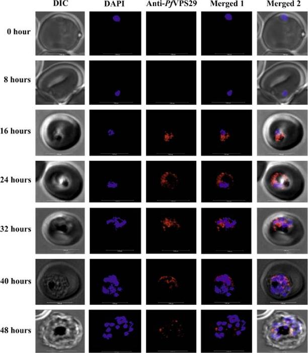
Localization of PfVPS29 in P. falciparum. Ring synchronized parasite (0 h) was grown in culture and at different time points of growth, iRBCs were processed and incubated with anti-PfVPS29 antibody followed by anti-rabbit Alexa fluor 647 (red fluorescence) and DAPI for nuclear staining (blue fluorescence). Each row is demonstrating a specific time point of growth from ring stage. Scale bar indicates 5 mm. PfVPS29 was located in the cytoplasm of the parasite. Although, PfVPS29 was clearly present from the trophozoite (16-32 h) up to the schizont (32-48 h) stage, its expression was observed maximally during the late trophozoite stage as evident from the fluorescent signal.Iqbal MS, Siddiqui AA, Alam A, Goyal M, Banerjee C, Sarkar S, Mazumder S, De R, Nag S, Saha SJ, Bandyopadhyay U. Expression, purification and characterization of Plasmodium falciparum vacuolar protein sorting 29. Protein Expr Purif. 2015 Dec 12. [Epub ahead of print]
See original on MMPMore information
| PlasmoDB | PF3D7_1406700 |
| GeneDB | PF3D7_1406700 |
| Malaria Metabolic Pathways | Localisation images Pathways mapped to |
| Previous ID(s) | PF14_0064 |
| Orthologs | PBANKA_1035500 , PCHAS_1036300 , PKNH_1351400 , PVP01_1342000 , PVX_086095 , PY17X_1037900 |
| Google Scholar | Search for all mentions of this gene |