PF3D7_1406300 glycerophosphodiester phosphodiesterase (GDPD)
Disruptability [+]
| Species | Disruptability | Reference | Submitter |
|---|---|---|---|
| P. falciparum 3D7 |
Refractory |
23000576 | Theo Sanderson, Wellcome Trust Sanger Institute |
| P. falciparum 3D7 |
Refractory |
USF piggyBac screen (Insert. mut.) | USF PiggyBac Screen |
Mutant phenotypes [+]
| Species | Stage | Phenotype | Reference | Submitter |
|---|---|---|---|---|
| P. falciparum 3D7 | Asexual |
Cell cycle arrest |
https://www.biorxiv.org/content/10.1101/2022.06.14.496138v1.full
(Conditional)
Growth defect at 24h trophozoite stages |
Abhinay Ramaprasad, |
Imaging data (from Malaria Metabolic Pathways)
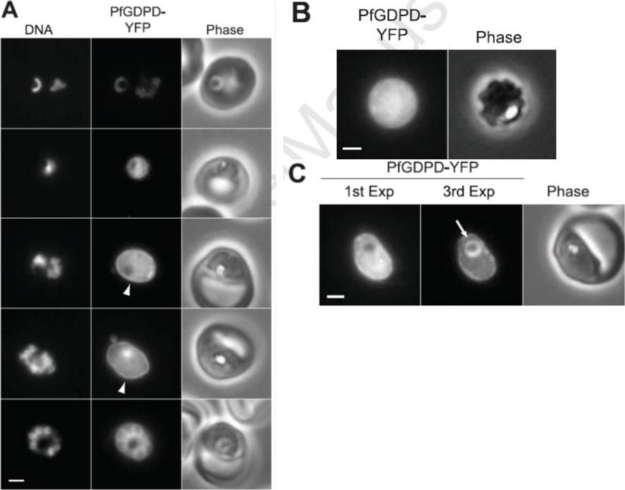
Localization of PfGDPD in asexual blood stage parasites. (A) Distribution of PfGDPD-YFP in live parasites at various stages of the asexual blood cycle. Parasites increase in maturity going from the top row (rings) to the bottom row (segmenting schizont). Rim fluorescence (PV) in mature trophozoites is indicated with an arrowhead. The exposure times and post acquisition processing parameters were identical for all YFP images to provide a sense of the relative expression levels of PfGDPD-YFP during the blood cycle. DNA was stained with Hoechst 33342. Bar, 2 mm. (B) Image of a parasite expressing PfGDPD-YFP that has been treated with 0.25 mg/mL saponin to permeabilize the erythrocyte and PV membranes. Bar, 2 mm. (C) The presence of YFP in the food vacuole is revealed after repeated exposures to excitation illumination. In the first one-second exposure (1st Exp), bright PV and cytosolic YFP fluorescence obscures the food vacuole fluorescence. Upon bleaching of PV and cytosolic YFP, the food vacuole fluorescence (arrow) becomes visible (3rd Exp). Bar, 2 mm.Denloye T, Dalal S, Klemba M. Characterization of a glycerophosphodiesterase with an unusual tripartite distribution and an important role in the asexual blood stages of Plasmodium falciparum. Mol Biochem Parasitol. 2012 186(1):29-37
See original on MMP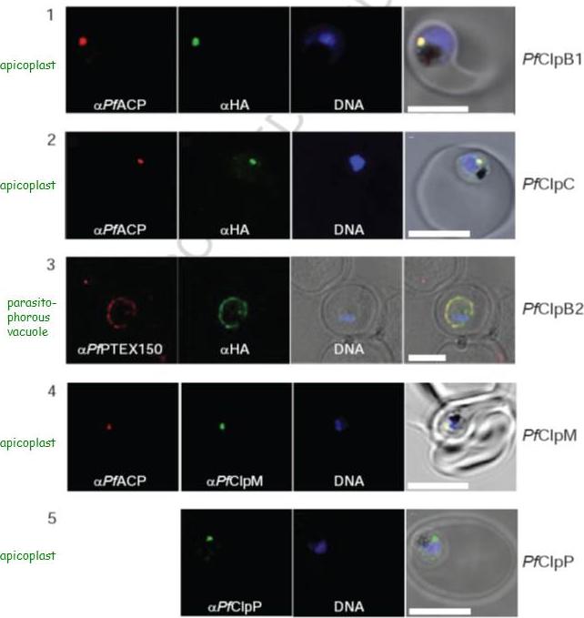
Localization of PfClpB1, PfClpC, PfClpB2, PfClpM, and PfClpP of P. falciparum was tested by immunofluorescent colocalization. In panels 1 and 2, PfClpB1-Strp-3×HA or PfClpC-Strp-3×HA parasites were probed with the apicoplast marker anti-ACP PFB0385w antisera (red), with anti-HA antisera (green), and with Hoescht 33342 to stain for DNA (blue). In panel 3, PfClpB2-Strp-3×HA parasites were probed with the parasitophorous vacuole marker anti-PTEX150 antisera (red), with anti-HA antisera (green), and with Hoescht 33342 (blue). In panels 4 and 5, wild type 3D7 strain was probed with anti-ACP antisera (red), with anti-PfClpM antisera or anti-ClpP antisera (green), and with Hoescht 33342 (blue). For all panels, the right most image represents the merged fluorescence images with the DIC or transmission image. The scale bars correspond to 5 mm.El Bakkouri M, Pow A, Mulichak A, Cheung KL, Artz JD, Amani M, Fell S, de Koning T, Goodman CD, McFadden GI, Ortega J, Hui R, Houry WA. The Clp chaperones and proteases of the human malaria parasite Plasmodium falciparum. J Mol Biol. 2010 404(3):456-77
See original on MMPMore information
| PlasmoDB | PF3D7_1406300 |
| GeneDB | PF3D7_1406300 |
| Malaria Metabolic Pathways | Localisation images Pathways mapped to |
| Previous ID(s) | PF14_0060 |
| Orthologs | PBANKA_1035900 , PCHAS_1036700 , PKNH_1351800 , PVP01_1342400 , PVX_086115 , PY17X_1038300 |
| Google Scholar | Search for all mentions of this gene |