PF3D7_1401800 choline kinase (CK)
Disruptability [+]
| Species | Disruptability | Reference | Submitter | |
|---|---|---|---|---|
| P. falciparum 3D7 |
Refractory |
USF piggyBac screen (Insert. mut.) | USF PiggyBac Screen | |
| P. berghei ANKA |
Refractory |
RMgm-818 | Imported from RMgmDB | |
| P. berghei ANKA |
Refractory |
RMgm-334 | Imported from RMgmDB | |
Mutant phenotypes [+]
| Species | Stage | Phenotype | Reference | Submitter |
|---|---|---|---|---|
| P. berghei ANKA | Asexual |
Attenuated |
PlasmoGEM (Barseq) | PlasmoGEM |
Imaging data (from Malaria Metabolic Pathways)
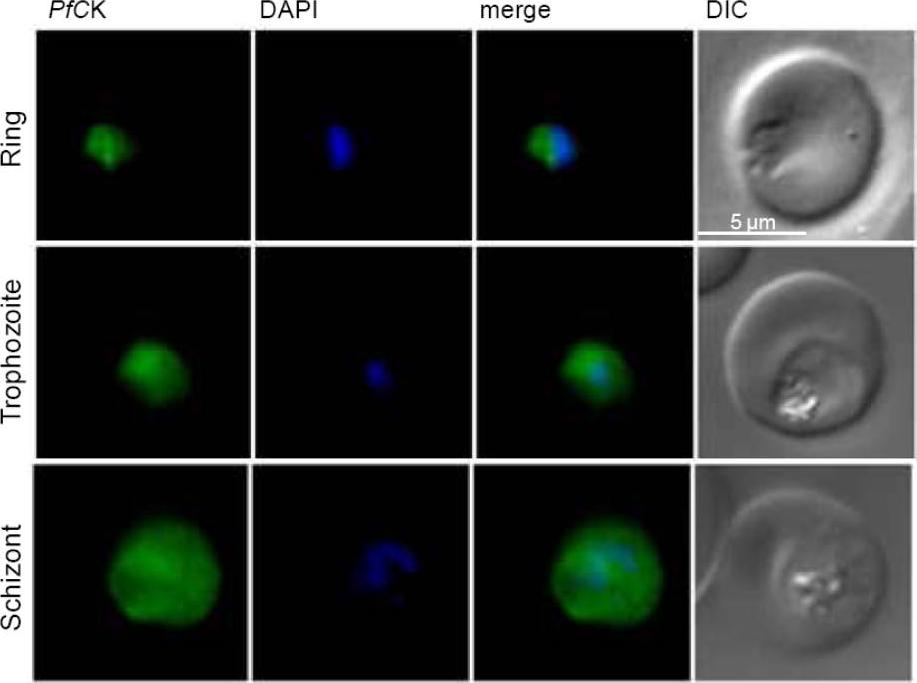
Immunofluorescence assays were done on human red blood cells infected by P. falciparum 3D7 strain and revealed by anti-PfCK. Nuclei were stained in blue with DAPI. Differential interference contrast (DIC) images and merges are presented. Three representative parasitic stages are shown: ring, mature trophozoite and schizont. The antibody was seen to be spread throughout the parasite cytoplasm.Alberge B, Gannoun-Zaki L, Bascunana C, Tran Van Ba C, Vial H, Cerdan R. Comparison of the cellular and biochemical properties of Plasmodium falciparum choline and ethanolamine kinases. Biochem J. 425:149-58, 2009. © The Biochemical Society 2009
See original on MMP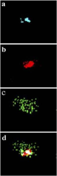
Confocal microscopy. Same field was observed with excitation wavelengths of 350 nm (DAPI), 579 nm (MitoTracker Red CMXRos) and 489 nm (Cy2). Blue, red and green fluorescence indicated the location of (a) nucleus (b) mitochondria and (c) PfCK, in Plasmodium falciparum, respectively. (d) Merged picture of above three individual pictures. The merged picture clearly suggested that PfCK is distributed throughout the cytoplasm (Fig. 6B, d) but not in the nucleus or mitochondria. Thus confocal studies further confirmed the cytosolic location of PfCK in P. falciparum.Choubey V, Guha M, Maity P, Kumar S, Raghunandan R, Maulik PR, Mitra K, Halder UC, Bandyopadhyay U. Molecular characterization and localization of Plasmodium falciparum choline kinase. Biochim Biophys Acta. 2006 1760:1027-38.
See original on MMP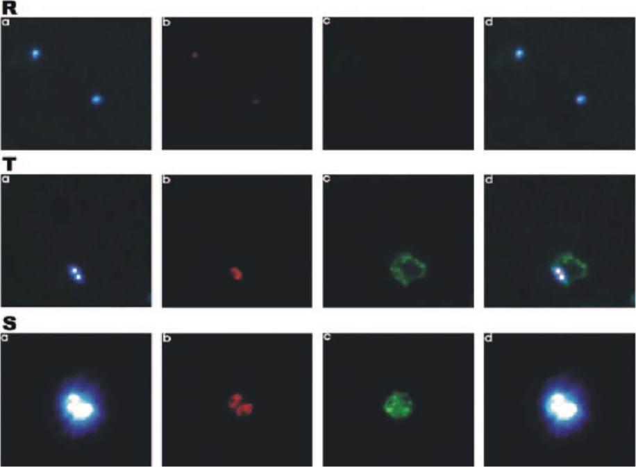
Stage-specific expression of PfCK by immunofluorescence microscopy. Different fields were observed with excitation wavelengths of 350 nm (DAPI), 579 nm (MitoTracker Red CMXRos), and 489 nm (Cy2). Blue, red, and green fluorescence indicate the locations of nucleus (a), mitochondria (b), and PfCK (c) in Plasmodium falciparum, respectively. (d) Merged picture of panel a, b, and c pictures.Choubey V, Maity P, Guha M, Kumar S, Srivastava K, Puri SK, Bandyopadhyay U. Inhibition of Plasmodium falciparum choline kinase by hexadecyltrimethylammonium bromide: a possible antimalarial mechanism. Antimicrob Agents Chemother. 2007 51:696-706.
See original on MMPMore information
| PlasmoDB | PF3D7_1401800 |
| GeneDB | PF3D7_1401800 |
| Malaria Metabolic Pathways | Localisation images Pathways mapped to |
| Previous ID(s) | PF14_0020 |
| Orthologs | PBANKA_1040100 , PCHAS_1040900 , PKNH_1356300 , PVP01_1347000 , PVX_086340 , PY17X_1042600 |
| Google Scholar | Search for all mentions of this gene |