PF3D7_1361900 proliferating cell nuclear antigen 1 (PCNA1)
Disruptability [+]
| Species | Disruptability | Reference | Submitter |
|---|---|---|---|
| P. falciparum 3D7 |
Refractory |
USF piggyBac screen (Insert. mut.) | USF PiggyBac Screen |
Mutant phenotypes [+]
None reported yet. Please press the '+' button above to add one.Imaging data (from Malaria Metabolic Pathways)
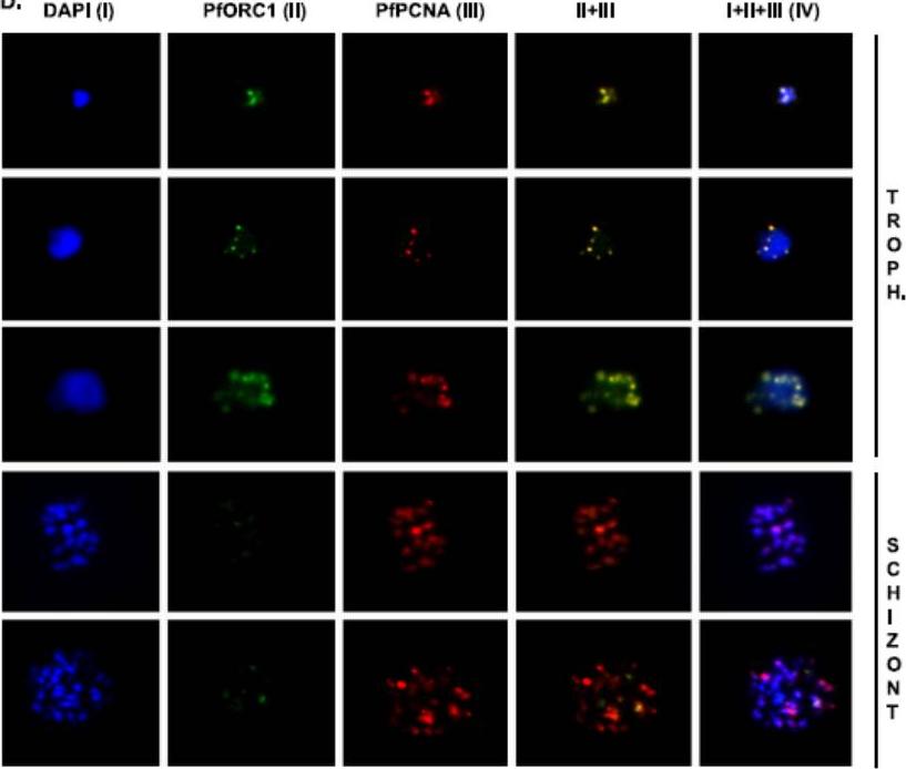
Colocalization of PfPCNA1 and PfORC1 during the replicating-trophozoite stage. A glass slide containing a parasite smear from the replicating-trophozoite stage or schizont stage was treated for immunofluoresence studies as described in Materials and Methods using both anti-PfORC1 and anti-PfPCNA1 antibodies. Distinct colocalization of PfORC1 and PfPCNA1 foci can be detected during the replicating-trophozoite stage (Troph.; rows 1 to 3). Although the expression of PfPCNA can be detected at the multinucleated late schizont stage (rows 4 and 5), the expression of PfORC1 is barely visible as the protein is degraded at the late stage, as described earlier. DAPI (4’,6-diamidino-2-phenylindole) (I) shows the nuclei. Column IV represents merged panels I, II, and III. Gupta A, Mehra P, Deshmukh A, Dar A, Mitra P, Roy N, Dhar SK. Functional dissection of the catalytic carboxyl-terminal domain of origin recognition complex subunit 1 (PfORC1) of the human malaria parasite Plasmodium falciparum. Eukaryot Cell. 2009 8:1341-51.
See original on MMP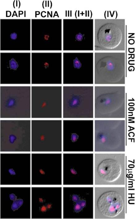
Effect of ACF on replication foci formation. Immunofluorescence assay to show pattern of replication foci (PCNA foci) formation during parasite developmental stages in untreated, Acriflavine (100 nM) and hydroxyurea (70 μg/mL) treated parasites. DAPI was used for nuclear staining. Panel III shows the merged images of DAPI (I) and PCNA (II) signals whereas panel IV represents the merged images of PCNA signal, DAPI and DIC images. Immunofluorescence results indicate the presence of diffused signals of PCNA in Acriflavine as well as hydroxyurea- treated parasites compared to the distinct nuclear foci found in untreated parasites. Dana S, Prusty D, Dhayal D, Gupta MK, Dar A, Sen S, Mukhopadhyay P, Adak T, Dhar SK. Potent antimalarial activity of acriflavine in vitro and in vivo. ACS Chem Biol. 2014 9(10):2366-73.
See original on MMP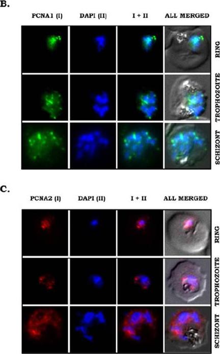
Stage-specific expression and subcellular localization of PfPCNA proteins. (B and C) IFA was performed on glass slides containing parasite smears, which were treated with antibodies against PfPCNA1 (B) or PfPCNA2 (C). PfPCNA1 (green) shows nuclear foci formation in replicating trophozoite stage and multinucleate schizont stage, whereas PfPCNA2 (red) shows both nuclear and cytoplasmic diffuse staining. DAPI (blue) stains the nuclei. The IFA showed distinct nuclear foci of endogenous PCNA1 during trophozoite and schizont stages of the parasites (B). Interestingly, endogenous PfPCNA2 shows a more diffused pattern throughout the nucleus and cytoplasm (C).Mitra P, Banu K, Deshmukh AS, Subbarao N, Dhar SK. Functional dissection of proliferating-cell nuclear antigens (1 and 2) in human malarial parasite Plasmodium falciparum: possible involvement in DNA replication and DNA damage response. Biochem J. 2015 470(1):115-29. PMID: 1.
See original on MMP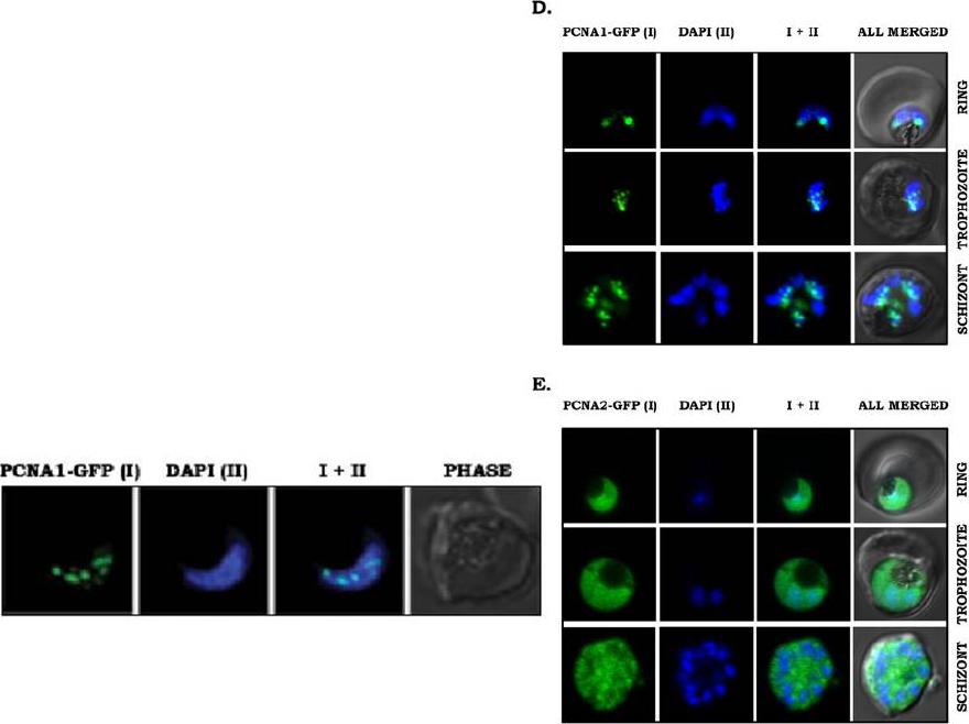
Right panels: Stage-specific expression and subcellular localization of PfPCNA proteins. (D and E) PfPCNA1–GFP and PfPCNA2–GFP transgenic parasite lines were visualized using an Olympus confocal microscope and Olympus Fluoview software. PfPCNA1–GFP (D) predominantly shows nuclear punctate staining co-localized with DAPI. PfPCNA2–GFP (E) shows a diffuse staining pattern across the parasite including the nuclei stained with DAPI. PCNA1–GFP showed distinct nuclear foci similar to endogenous protein (2D). However, these foci were absent from PCNA2–GFP, where a diffuse staining pattern could be seen throughout the parasites with slight enrichment of PCNA2–GFP in the nucleus (E). :pwer left panel: PCNA1 marks the sites of active replication. Live cell imaging of PCNA1-GFP expressing parasites using high resolution confocal microscopy shows distinct PCNA1 foci associated with the nucleus (shown in blue, DAPI) in trophozoite stages of the parasites.Mitra P, Banu K, Deshmukh AS, Subbarao N, Dhar SK. Functional dissection of proliferating-cell nuclear antigens (1 and 2) in human malarial parasite Plasmodium falciparum: possible involvement in DNA replication and DNA damage response. Biochem J. 2015 470(1):115-29.
See original on MMPMore information
| PlasmoDB | PF3D7_1361900 |
| GeneDB | PF3D7_1361900 |
| Malaria Metabolic Pathways | Localisation images Pathways mapped to |
| Previous ID(s) | PF13_0328 |
| Orthologs | PBANKA_1137900 , PCHAS_1137400 , PKNH_1109400 , PVP01_1110100 , PVX_115055 , PY17X_1139300 |
| Google Scholar | Search for all mentions of this gene |