PF3D7_1359400 CUGBP Elav-like family member 1 (CELF1)
Disruptability [+]
No information reported yet. Please press the '+' button above to add some.Mutant phenotypes [+]
None reported yet. Please press the '+' button above to add one.Imaging data (from Malaria Metabolic Pathways)
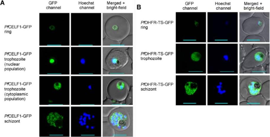
Protein localization of PfCELF1 and PfDHFR-TS. PfCELF1 was expressed as a fusion protein with C-terminal GFP in a transgenic parasite line established in 3D7 strain. The integrant transgenic line clone #1 expressing GFP-tagged PfDHFR-TS established in 3D7 strain for comparison was described previously [36]. (A) Representative confocal microscopic images of transgenic PfCELF1-GFP parasites. Images are shown for each asexual developmental stage (ring, trophozoite and schizont); left panel is the GFP signal showing PfCELF1-GFP, middle panel is Hoechst signal showing nucleus and right panel is the merge of both fluorescent signals overlaid with the bright-field image. Scale bars, 5 mm. (B) Representative confocal microscopic images of transgenic PfDHFR-TS-GFP parasites. Images are shown for each asexual developmental stage and arranged as in part A. Scale bars, 5 mm. Wongsombat C, Aroonsri A, Kamchonwongpaisan S, Morgan HP, Walkinshaw MD, Yuthavong Y, Shaw PJ. Molecular characterization of Plasmodium falciparumBruno/CELF RNA binding proteins. Mol Biochem Parasitol. 2014 Nov 3;198(1):1-10. [Epub ahead of print]
See original on MMPMore information
| PlasmoDB | PF3D7_1359400 |
| GeneDB | PF3D7_1359400 |
| Malaria Metabolic Pathways | Localisation images Pathways mapped to |
| Previous ID(s) | PF13_0315 |
| Orthologs | PBANKA_1135700 , PCHAS_1135200 , PKNH_1111900 , PVP01_1112500 , PVX_114940 , PY17X_1137200 |
| Google Scholar | Search for all mentions of this gene |