PF3D7_1353200 membrane associated histidine-rich protein (MAHRP2)
Disruptability [+]
| Species | Disruptability | Reference | Submitter |
|---|---|---|---|
| P. falciparum 3D7 |
Refractory |
20624222 | Theo Sanderson, Wellcome Trust Sanger Institute |
| P. falciparum 3D7 |
Possible |
USF piggyBac screen (Insert. mut.) | USF PiggyBac Screen |
Mutant phenotypes [+]
None reported yet. Please press the '+' button above to add one.Imaging data (from Malaria Metabolic Pathways)

A. tage dependent co-localization of PfHsp70-x and PfEMP1. Upper infected cell (ring stage) shows a higher degree of signal colocalization than lower infected cell (trophozoite). B. ndirect immuno-fluorescence of PfHsp70-x and PfEMP1 using confocal microscopy. In merge and overlay: Green, α-70x; Red, α-ATS; Blue, Hoechst. C. ndirect immuno-fluorescence localization of PfHsp70-x using anti-SBP1, anti-MAHRP2 and anti-STEVOR (STV) antisera in cells infected with 3D7GFP:70x. In merge and overlay images: Green, GFP; red, specific antisera; Blue, Hoechst. No co-localization can be observed between the GFP and antibody-specific signals.Külzer S, Charnaud S, Dagan T, Riedel J, Mandal P, Pesce ER, Blatch GL, Crabb BS, Gilson PR, Przyborski JM. Plasmodium falciparum-encoded exported hsp70/hsp40 chaperone/co-chaperone complexes within the host erythrocyte. Cell Microbiol. 2012 14(11):1784-95
See original on MMP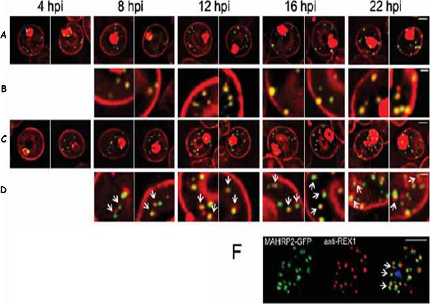
Live cell time course showing assembly of exomembrane components. Transfectants expressing GFP chimeras of (A, B) REX1 and (C, D) MAHRP2 were co-labeled with BODIPYceramide. Infected RBCs were synchronized to a ~2 h window and samples were collected at 4 h intervals. BODIPY-ceramide- and REX1-GFP-labeled Maurer's clefts and MAHRP2-GFP-labled structures are evident in the RBC cytoplasm from ~4 h post invasion. MAHRP2-GFP-labled structures are associated with the BODIPY-ceramide-labeled Maurer's clefts (D, white arrows). (F) MAHRP2-GFP transfectants were permeabilized with EqtII and labeled with antibodies recognizing REX1. Scale bars = 3 μm (A, C), 1 μm (B, D) and 4 μm (F). McMillan PJ, Millet C, Batinovic S, Maiorca M, Hanssen E, Kenny S, Muhle RA, Melcher M, Fidock DA, Smith JD, Dixon MW, Tilley L. Spatial and temporal mapping of the PfEMP1 export pathway in Plasmodium falciparum. Cell Microbiol. 2013 15(8):1401-18.
See original on MMP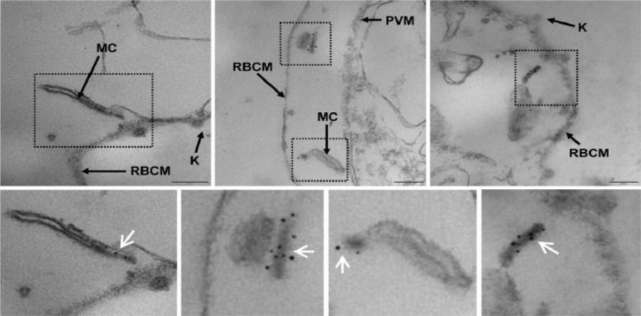
MAHRP2 is associated with electron-dense tubules in the cytoplasm of infected erythrocytes. Thin sections through EqtII-permeabilized erythrocytes infected with 3D7 parasites. The sections were probed with rabbit anti-MAHRP2 antibody, and subsequently decorated with protein A-gold conjugate (6 nm). Maurer’s clefts (MC) have an electron-lucent lumen and an electron-dense coat. The structures labelled by MAHRP2 are slender electron-dense tubules (white arrows) that are often associated with the Maurer’s clefts. K, Knobs; RBCM, red blood cell membrane; PVM, PV membrane. Bars are 200 nm.Pachlatko E, Rusch S, Müller A, Hemphill A, Tilley L, Hanssen E, Beck HP. MAHRP2, an exported protein of Plasmodium falciparum, is an essential component of Maurer's cleft tethers. Mol Microbiol. 2010 77(5):1136-52
See original on MMP
Dual label immunofluorescence microscopy of formaldehyde or acetone/methanol-fixed 3D7 smears using antibodies recognizing MAHRP2 (green) and anti-MAHRP1, anti-SBP1 and anti-PfEMP1 (red), or (last panel) anti-MAHRP1 (green) and anti-SBP1 (red). Scale bar = 5 mm. In mature stage parasites the antibodies reacted with punctate structures in the erythrocyte cytoplasm in a pattern that is somewhat reminiscent of Maurer’s cleft staining. No colocalization of MAHRP2 could be found with MAHRP1 MAL13P1.413, SBP1 PFE0065w or PfEMP1; however, the signals were often in close proximity. This suggests that MAHRP2 is associated with a separate compartment of the exomembrane system of P. falciparum.Pachlatko E, Rusch S, Müller A, Hemphill A, Tilley L, Hanssen E, Beck HP. MAHRP2, an exported protein of Plasmodium falciparum, is an essential component of Maurer's cleft tethers. Mol Microbiol. 2010 77(5):1136-52
See original on MMP
Parasites were tagged by single cross-over homologous recombination, the endogenous gene locus to include the GFP coding sequence. PFE55INT. Co-immunofluorescence analysis on erythrocytes infected with PFE55INT, using anti-sera directed against STEVOR (A), MAHRP1 MAL13P1.413 (B), KAHRP PFB0100c (C). Fluorescence channels are shown individually in black/white for highest contrast. All images are maximal projections of Z-stack serial sections. In merge image green, GFP; red, STEVOR (A), MAHRP (B), KAHRP (C) or ATS domain of PfEMP1 (D); blue, Hoechst. Inset shows enlargement of merge (white box). The fusion protein labelled punctate structures within the infected erythrocytebut clearly not in Maurer’s clefts.Külzer S, Rug M, Brinkmann K, Cannon P, Cowman A, Lingelbach K, Blatch GL, Maier AG, Przyborski JM. Parasite encoded Hsp40 proteins define novel mobile structures in the cytosol of the P. falciparum infected erythrocyte. Cell Microbiol. 2010 12(10):1398-420 Copyright John Wiley & Sons Ltd. 2010.
See original on MMP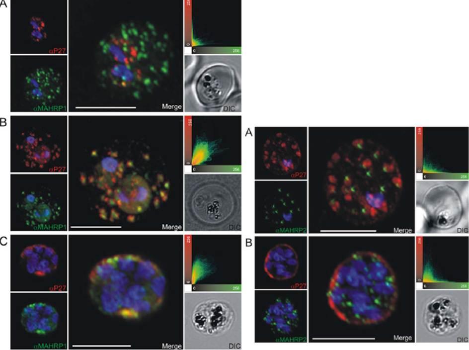
Left: Co-localization of Tex1 with MAHRP1. P27-specific polyclonal rabbit sera was used to detect Tex1 (red). Co-localization was performed using MAHRP1 polyclonal mouse sera (green). Co-localization was performed in ring stage (A) trophozoite (B) and schizont stage (C) infected RBC. Nuclear DNA was stained with DAPI (blue), Transmission image (DIC), Scale bar: 5 mm.Right: Tex1 localization in trophozoite and schizont stages with respect to newly described structures called tethers. Colocalization of Tex1 (red) with MAHRP2 (green) A) in trophozoite stages and B) in schizont stages. Nuclear DNA was stained with DAPI (blue), Transmission image (DIC), Scale bar: 5 mm.Kulangara C, Luedin S, Dietz O, Rusch S, Frank G, Mueller D, Moser M, Kajava AV, Corradin G, Beck HP, Felger I. Cell biological characterization of the malaria vaccine candidate trophozoite exported protein 1. PLoS One. 2012;7(10):e46112.
See original on MMP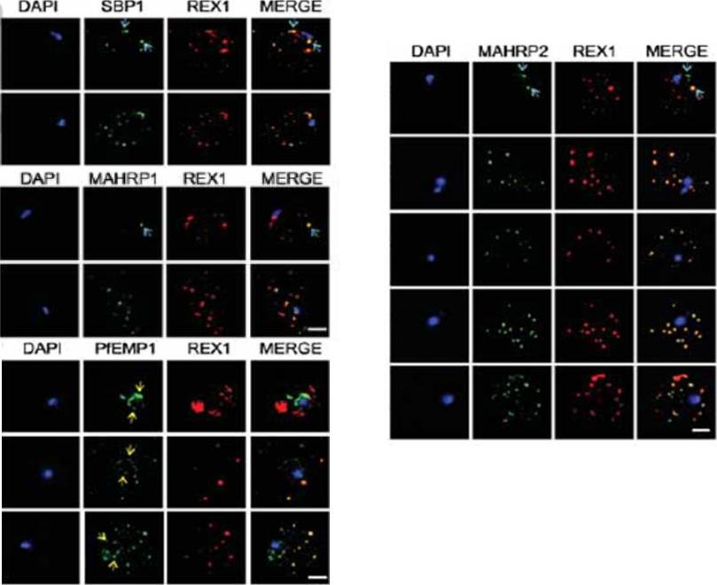
Immunofluorescence microscopy showing staggered delivery of different exomembrane components. (A-D) Infected RBCs were synchronized to a 1 h window, samples were collected at 2 h intervals and smears were fixed with acetone and stained with antibodies recognizing REX1, MAHRP1, SBP1, MAHRP2 and PfEMP1 (ATS) and co-stained with DAPI. Images are presented at time points before and after delivery to the RBC cytoplasm. Scale bars = 3 μm. McMillan PJ, Millet C, Batinovic S, Maiorca M, Hanssen E, Kenny S, Muhle RA, Melcher M, Fidock DA, Smith JD, Dixon MW, Tilley L. Spatial and temporal mapping of the PfEMP1 export pathway in Plasmodium falciparum. Cell Microbiol. 2013 15(8):1401-18
See original on MMP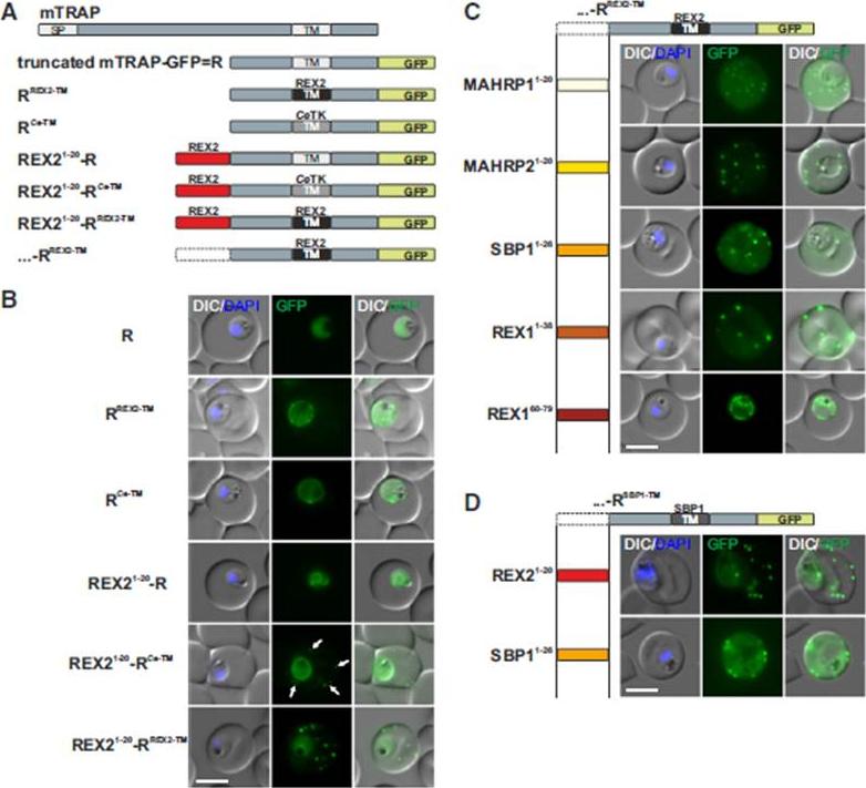
N Termini of PNEPs Are Sufficient for Export of RREX2-TM, (A) Schematic of mTRAP fusion constructs. (B) Representative images of live P. falciparum parasites expressing the constructs shown in (A). DIC, differential interference contrast; nuclei were stained with DAPI. Arrows indicate limited staining reminiscent of Maurer’s clefts. (C and D) Images of live P. falciparum parasites expressing RREX2-TM (C) and RSBP1-TM (D) fused with the PNEP N termini indicated. Panels are as in (B). Size bars represent 5 mm. The mTRAP backbone, different TMs, and the SP (signal peptide) are shown in different shades of gray; N-terminally appended regions of PNEPs are shown in shades from yellow to dark red. Ce-TM, C. elegans TK TM. The unmodified truncated mTRAP fused to GFP (‘‘R’’ for ‘‘reporter’’) does not contain any export-relevant sequences. This protein was found evenly distributed in the parasite cytosol (B). When the mTRAP TM in R was replaced with that of the PNEP REX2 or a previously used heterologous TM of a C.elegans tyrosine kinase (TK) (a TM that does not promote export of REX2 and does not change topology;), the R then entered the secretory pathway and was found in the parasite periphery (PPM, PV, or PVM) but was not exported.Grüring C, Heiber A, Kruse F, Flemming S, Franci G, Colombo SF, Fasana E,Schoeler H, Borgese N, Stunnenberg HG, Przyborski JM, Gilberger TW, Spielmann T. Uncovering common principles in protein export of malaria parasites. Cell Host Microbe. 2012 12(5):717-29.
See original on MMP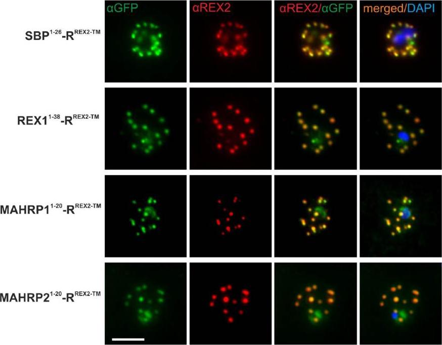
Immunofluorescence Assays Confirm Maurer’s Clefts Localization of PNEP N Termini mTRAP Reporter Fusions. Acetone fixed parasites expressing the reporter indicated on the left were probed with anti-GFP and a serum reacting with the C-terminus of REX2. Under these fixing conditions soluble proteins in the host cell are lost, leading to absence of the soluble pool of the reporter. DIC, differential interference contrast; DAPI, nuclei. Size bar: 5m.Grüring C, Heiber A, Kruse F, Ungefehr J, Gilberger TW, Spielmann T. Development and host cell modifications of Plasmodium falciparum blood stages in four dimensions. Nat Commun. 2011 2:165. PMID:
See original on MMP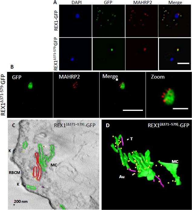
Analysis of the Maurer’s clefts ultrastructure and distribution of tethers ofREX1(Δ371–579)-GFP parasites. REX1-GFP and REX1(Δ371–579)-GFP transfectants were fixed with paraformaldehyde/ glutaraldehyde, permeabilized with Triton X-100, and probed with anti-GFP (green) and anti-MAHRP2 (red). Samples imaged using (A) widefield deconvolution microscopy or (B) 3D-SIM. Scale bars = 3 μm; zoom bar = 1 μm. C. STEM tomogram (600 nm section) of an EqII-permeabilized REX1(Δ371–579)-GFP-infected RBC showing the stacked Maurer’s clefts (MC) layers, and knobs (K) on the RBC membrane (RBCM). The lamella indicated in red share a membrane continuum. D. Rendered STEM tomogram of REX1(Δ371–579)-GFP-infected RBC labelled with anti-GFP antibodies and protein A gold showing Maurer’s clefts (MC, green), tethers (T, magenta) and gold particles (Au, yellow). In REX1-GFP parasites, immunofluorescence reveals MAHRP2 labelling (Fig. 6A) closely adjacent to the REX1-GFP labelled Maurer's clefts. For the REX1Δ371-579-GFP parasites, the MAHRP2 labelling partly overlaps with and partly sits outside the single puncta of GFP labelling. McHugh E, Batinovic S, Hanssen E, McMillan PJ, Kenny S, Griffin MD, Crawford S, Trenholme KR, Gardiner DL, Dixon MW, Tilley L. A repeat sequence domain of the ring-exported protein-1 of Plasmodium falciparum controls export machinery architecture and virulence protein trafficking. Mol Microbiol. 2015 Aug 24. [Epub ahead of print]
See original on MMPMore information
| PlasmoDB | PF3D7_1353200 |
| GeneDB | PF3D7_1353200 |
| Malaria Metabolic Pathways | Localisation images Pathways mapped to |
| Previous ID(s) | PF13_0276 |
| Orthologs | |
| Google Scholar | Search for all mentions of this gene |