PF3D7_1345100 thioredoxin 2 (TRX2)
Disruptability [+]
| Species | Disruptability | Reference | Submitter | |
|---|---|---|---|---|
| P. falciparum 3D7 |
Refractory |
USF piggyBac screen (Insert. mut.) | USF PiggyBac Screen | |
| P. berghei ANKA |
Possible |
RMgm-943 | Imported from RMgmDB | |
| P. berghei ANKA |
Possible |
RMgm-918 | Imported from RMgmDB | |
| P. berghei ANKA |
Possible |
PlasmoGEM (Barseq) | PlasmoGEM | |
Mutant phenotypes [+]
| Species | Stage | Phenotype | Reference | Submitter |
|---|---|---|---|---|
| P. berghei ANKA | Asexual |
Difference from wild-type |
RMgm-943
Reduced asexual multiplication/growth rate |
Imported from RMgmDB |
| P. berghei ANKA | Asexual |
Difference from wild-type |
RMgm-918
Blood stages show a delayed growth in mice. Evidence is presented for a prolonged cell cycle of asexual blood stages, mainly affecting the development of ring forms and schizonts. The number of merozoites per schizont was comparable to that of wild type schizonts. |
Imported from RMgmDB |
| P. berghei ANKA | Asexual |
Attenuated |
PlasmoGEM (Barseq) | PlasmoGEM |
| P. berghei ANKA | Gametocyte |
No difference |
RMgm-943 | Imported from RMgmDB |
| P. berghei ANKA | Oocyst |
No difference |
RMgm-943 | Imported from RMgmDB |
| P. berghei ANKA | Sporozoite |
No difference |
RMgm-943 | Imported from RMgmDB |
| P. berghei ANKA | Liver |
No difference |
RMgm-943 | Imported from RMgmDB |
Imaging data (from Malaria Metabolic Pathways)
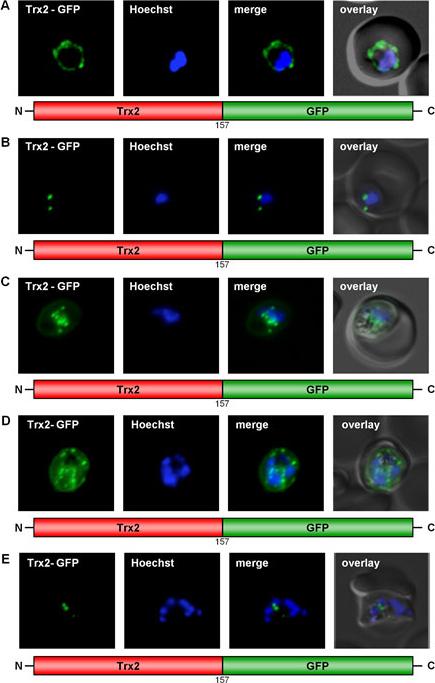
GFP targeting by thioredoxin 2 in various stages of P. falciparum. (A) Early ring stage parasite showing Trx2-GFP targeting to the PV. (B) Early trophozoite stage parasite showing two fluorescent Trx2-GFP points lying directly under the parasite plasma membrane. (C) Late trophozoite/early schizont stage parasite showing numerous fluorescent Trx2-GFP points. (D) Schizont stage parasite showing numerous fluorescent Trx2-GFP points. (E) Schizont stage parasite showing a small number of observable fluorescent Trx2-GFP structures. Live cell imaging of erythrocytes infected with transgenic parasites.Kehr S, Sturm N, Rahlfs S, Przyborski JM, Becker K. Compartmentation of redox metabolism in malaria parasites. PLoS Pathog. 2010 6:e1001242.
See original on MMP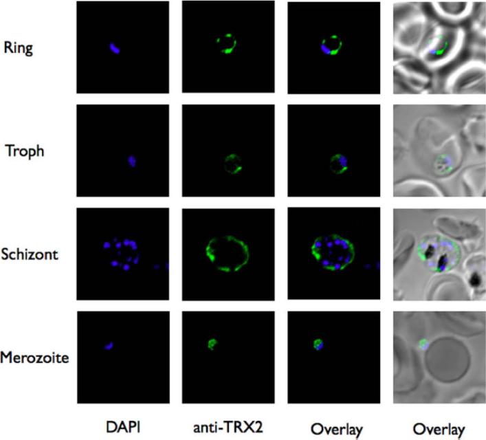
Visible and fluorescence images of 3D7 P. falciparum parasite immunostained with PfTrx-2 antibodies. PfTrx-2 is localized to the PVM (only) in different asexual stages of the parasite.Sharma A, Sharma A, Dixit S, Sharma A. Structural insights into thioredoxin-2: a component of malaria parasite protein secretion machinery. Sci Rep. 011;1:179.
See original on MMP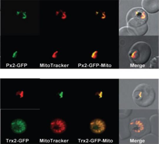
Localization of PfTrx-Px2 and PfTrx2 in P. falciparum erythrocytic stages. P. falciparum erythrocytic stages were transfected with construct pHH2-Px2-GFP, pHH2-Trx2-GFP leading to the expression of the peroxiredoxin or thioredoxin2 C-terminally fused to green fluorescent protein (GFP).A. The localization of the peroxiredoxin-GFP fusion protein in parasites previously treated with MitoTracker CMX-Ros was analysed by fluorescence light microscopy. Phase, phase contrast of parasitized erythrocytes infected with P. falciparum trophozoites; PfTrx-Px2-GFP, parasitized erythrocytes expressing the peroxiredoxin-GFP fusion protein analysed using the FITC channel; MitoTracker, parasitized erythrocytes expressing the peroxiredoxin-GFP fusion protein analysed using the rhodamine channel; PfTrx-Px2-Mito; merge of FITC and rhodamine channels; merge, merge of all images. The images show that the peroxiredoxin-GFP fusion protein is colocalizing with the mitochondrion (stained by MitoTracker).B. The expression of pHH2-PfTrx2 results in the localization of the fusion protein the mitochondrion. Phase, phase contrast of parasitized erythrocytes infected with P. falciparum trophozoites; PfTrx2-GFP, parasitized erythrocytes expressing the Trx2-GFP fusion protein analysed using the FITC channel; MitoTracker, parasitized erythrocytes expressing the Trx2-GFP fusion protein analysed using the rhodamine channel; PfTrx2-Mito; merge of FITC and rhodamine channels; merge, merge of all images.Boucher IW, McMillan PJ, Gabrielsen M, Akerman SE, Brannigan JA, Schnick C, Brzozowski AM, Wilkinson AJ, Müller S. Structural and biochemical characterization of a mitochondrial peroxiredoxin from Plasmodium falciparum. Mol Microbiol. 2006 61:948-59.PMID
See original on MMP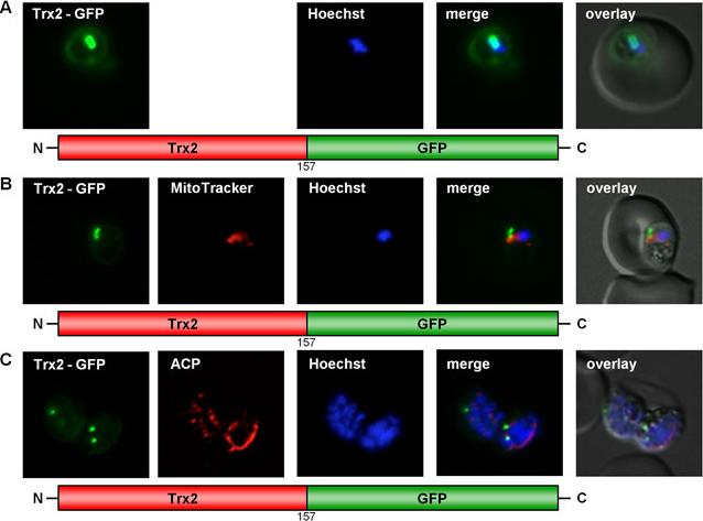
GFP targeting by P. falciparum thioredoxin 2. (A)–(C) Targeting of Trx2 to the parasitophorous vacuole and to a not yet characterized, non-dividing organelle within the parasite. Colocalization of GFP with the mitochondrial stain MitoTrackerOrange in fixed cells. Colocalization of GFP and the apicoplast marker ACP in fixed, immunodecorated cells. Trx2 is clearly localized in the parasitophorous vacuole. Trx2-GFP labeled further structures within the parasite (B, C) in .90% of the parasites. In these parasites, the GFP fluorescence did neither colocalize with the ER nor with the nucleus, the mitochondrion or the apicoplast, as evidenced by analysis using organellar markers.Kehr S, Sturm N, Rahlfs S, Przyborski JM, Becker K. Compartmentation of redox metabolism in malaria parasites. PLoS Pathog. 2010 6:e1001242.
See original on MMPMore information
| PlasmoDB | PF3D7_1345100 |
| GeneDB | PF3D7_1345100 |
| Malaria Metabolic Pathways | Localisation images Pathways mapped to |
| Previous ID(s) | MAL13P1.225 |
| Orthologs | PBANKA_1358000 , PCHAS_1362600 , PKNH_1256200 , PVP01_1209700 , PVX_083155 , PY17X_1363700 |
| Google Scholar | Search for all mentions of this gene |