PF3D7_1335400 reticulocyte binding protein 2 homologue a (RH2a)
Disruptability [+]
| Species | Disruptability | Reference | Submitter |
|---|---|---|---|
| P. falciparum 3D7 |
Possible |
19400777 | Theo Sanderson, Wellcome Trust Sanger Institute |
| P. falciparum 3D7 |
Possible |
USF piggyBac screen (Insert. mut.) | USF PiggyBac Screen |
Mutant phenotypes [+]
| Species | Stage | Phenotype | Reference | Submitter |
|---|---|---|---|---|
| P. falciparum 3D7 | Asexual |
Attenuated |
19400777 | Theo Sanderson, Wellcome Trust Sanger Institute |
Imaging data (from Malaria Metabolic Pathways)
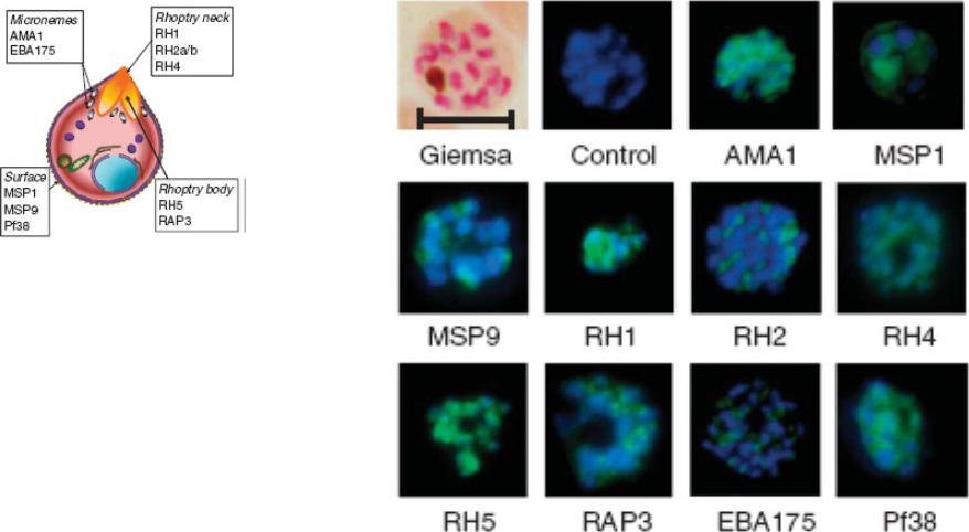
Indirect immunofluorescence images of P. falciparum schizonts stained with rabbit IgG (green) induced by 10 viral-vectored vaccines expressing malaria antigens, and negative control vectors lacking a malaria antigen. Nuclei are counterstained with 4,6-diamidino-2-phenylindole (blue). All sera were tested against 3D7 strain parasites, with the exception of anti-PfRH1 for which FVO strain parasites were used. All images to same scale as Giemsa stained image (top left, on which scale bar indicates 5 μm).Douglas AD, Williams AR, Illingworth JJ, Kamuyu G, Biswas S, Goodman AL, Wyllie DH, Crosnier C, Miura K, Wright GJ, Long CA, Osier FH, Marsh K, Turner AV, Hill AV, Draper SJ. The blood-stage malaria antigen PfRH5 is susceptible to vaccine-inducible cross-strain neutralizing antibody. Nat Commun. 2011 2:601.
See original on MMP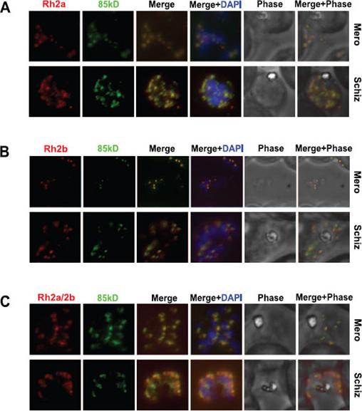
The 85 kDa processed form of PfRh2a and PfRh2b colocalises with the corresponding C-terminal regions in both merozoite and schizont stages. (A) The 85 kDa product co-localises with the C-terminal PfRh2a product. Free merozoites or schizonts of the 3D7 parasite were dual stained with 8F9 anti-PfRh2a and R1170 anti-85 kDa antibodies. Both merozoites and schizonts give an apical staining pattern with both antibodies, indicating co-localisation. Nuclei were stained with DAPI. (B) The 85 kDa product co-localises with the C-terminal PfRh2b product. Parasites were dual stained with 4B7 anti-PfRh2b and R1170 anti-85 kDa antibodies. As for (A), both antibodies give a co-localised apical staining pattern. (C) The 85 kDa product co-localises with both the PfRh2a and PfRh2b C-terminal products. Parasites were dual stained with 3A2 anti-PfRh2a/2b and R1170 anti-85 kDa antibodies. As for (A), both antibodies give a co-localised apical staining pattern.Triglia T, Chen L, Lopaticki S, Dekiwadia C, Riglar DT, Hodder AN, Ralph SA, Baum J, Cowman AF. Plasmodium falciparum merozoite invasion is inhibited by antibodies that target the PfRh2a and b binding domains. PLoS Pathog. 2011 Jun;7(6):e1002075.
See original on MMP
Subcellular localization of PfRh2a/b in 3D7 parasites by immunoelectron microscopy. (A-C) Cross-sections through merozoites. Scale bars are 200 nm in all panels. Immunoelectron microscopy with anti-PfRh2a/b antibodies showed labeling in the electron-dense rhoptry organelles of the parasite (A and C), with label evident in the neck of the rhoptry in some parasites (B).Duraisingh MT, Triglia T, Ralph SA, Rayner JC, Barnwell JW, McFadden GI, Cowman AF. Phenotypic variation of Plasmodium falciparum merozoite proteins directs receptor targeting for invasion of human erythrocytes. EMBO J. 2003 22:1047-57.
See original on MMP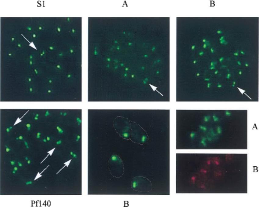
Immunofluorescence assays performed with antisera against fragments of the shared domain (S1), fragments of the unique regions, anti-PfRBP2-Ha (A) and anti-PfRBP2-Hb (B), and the rhoptry antigen Pf-140, on air-dried, acetone-fixed, P. falciparum-infected erythrocytes. The lower center panel (B) is at a higher magnification (33,750; others are 31,250), with the outline of the merozoites marked. The lower right shows costaining of single merozoites with rabbit anti-A serum (A) and rat anti-B serum (B). Each of the PfRBP2-H antisera, whether raised against fragments of the shared domain, anti-S1 and anti-S3, or against the unique regions, anti-A and anti-B, yielded a punctate staining pattern, generally with a single dot of fluorescence, although occasionally two foci were visible in a single merozoite (marked by arrows Upper). This double-dot pattern is reminiscent of proteins such as the Pf140 protein, which are localized to the paired rhoptry organelles. Higher magnification (Lower Center) reveals that the staining is concentrated at the apical end of the merozoites.Rayner JC, Galinski MR, Ingravallo P, Barnwell JW. Two Plasmodium falciparum genes express merozoite proteins that are related to Plasmodium vivax and Plasmodium yoelii adhesive proteins involved in host cell selection and invasion. Proc Natl Acad Sci U S A. 2000 97:9648-53. Copyright 2009 National Academy of Sciences, U.S.A.
See original on MMP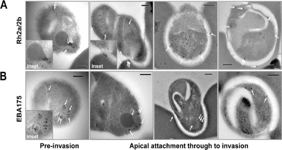
Immuno-electron microscopy localisation of PfRh2a and PfRh2b during merozoite invasion of human erythrocytes. (A) The PfRh2a/2b proteins localise to the rhoptry neck and moving junction of merozoites. Transmission electron micrographs of pre- and mid-invasion merozoites showing PfRh2a/b-specific immunogold labelling (white arrows). At the pre-invasion stage, PfRh2a/b localises to the rhoptry neck (inset), while during mid-invasion, PfRh2a/b localises both to the moving junction and to the merozoite surface within the invasion pit. Scale bar = 0.2 mm. (B) The EBA-175 protein localises to the micronemes and apical tip of merozoites. At the pre-invasion stage, EBA-175 is located within micronemes (inset), while during mid-invasion, EBA-175 can be located either at the apical tip or within the invasion pit. Scale bar = 0.2 mm.Triglia T, Chen L, Lopaticki S, Dekiwadia C, Riglar DT, Hodder AN, Ralph SA, Baum J, Cowman AF. Plasmodium falciparum merozoite invasion is inhibited by antibodies that target the PfRh2a and b binding domains. PLoS Pathog. 2011 (6):e1002075.
See original on MMP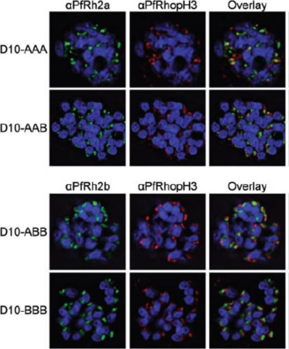
Chimeric protein localization and processing. D10-AAA, D10-AAB, D10-ABB and D10-BBB schizonts were methanol-fixed and stained with rabbit anti-PfRh2a or rabbit anti-PfRh2b, mouse anti-PfRhopH3 and DAPI. Images were obtained using deconvolution microscopy. The chimeric proteins from both D10-AAA and D10-AAB localized to the apical organelles (Fig. 3A, left panel) and in close proximity to PfRhopH3 (middle panel). Fluorescence overlays showed some direct colocalization between the PfRh2a/b proteins and PfRhopH3 (right panel). PfRh2a and PfRh2b are known to localize to the rhoptry neck whereas PfRhopH3 localizes to the rhoptry bulb.Dvorin JD, Bei AK, Coleman BI, Duraisingh MT. Functional diversification between two related Plasmodium falciparum merozoite invasion ligands is determined by changes in the cytoplasmic domain. Mol Microbiol. 2010 75:990-1006.
See original on MMP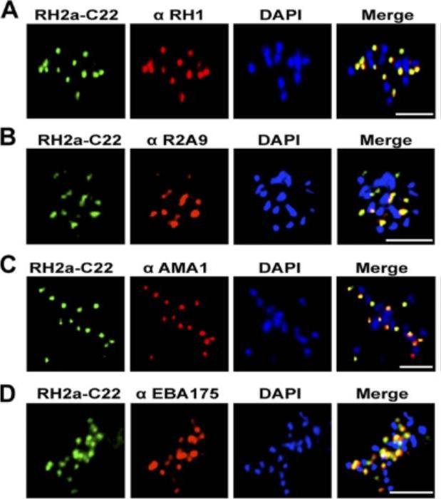
An immunofluorescence assay was performed on late-schizont-stage W2mef parasites stained for RH2a using the anti-RH2a MAb C22 and costained either with an antibody against the rhoptry neck marker RH1 (A), rabbit polyclonal antibody R2A9, recognizing both RH2a and RH2b (B), or an antibody against the micronemal marker AMA1 (C) or EBA175 (D). Bars, 5 mm. PfRH2a showed a punctate pattern at the apical tip of the merozoite in schizonts and appeared to colocalize with PfRH1 in W2mef and 3D7 parasites, strongly suggesting that RH2a is located at the rhoptry neck.Gunalan K, Gao X, Liew KJ, Preiser PR. Differences in erythrocyte receptor specificity of different parts of the Plasmodium falciparum reticulocyte binding protein homologue 2a. Infect Immun. 2011 79:3421-30.
See original on MMP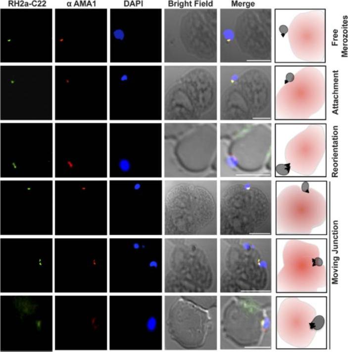
RH2a follows the moving junction during invasion. RH2a was costained with AMA1. From top to bottom, immunofluorescence images show merozoites progressing through different stages of invasion: free merozoites, an attached merozoite, a reoriented merozoite whose apical end is facing the red blood cells, and the progression of the moving junction. Bars, 5 mm.Gunalan K, Gao X, Liew KJ, Preiser PR. Differences in erythrocyte receptor specificity of different parts of the Plasmodium falciparum reticulocyte binding protein homologue 2a. Infect Immun. 2011 79:3421-30. PMID:
See original on MMP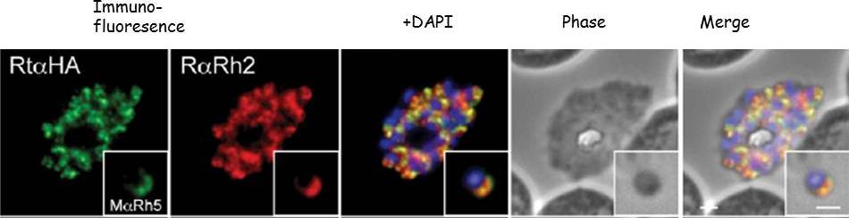
Immunofluorescence and phase contrast images of late schizonts or free merozoites (insets) to colocalise PfRh5 with PfRh2a/b. Each panel from left top right corresponds to anti-HA antibodies (to detect PhRh5), rabbit anti-PfRh2a/b, overlay of each with DAPI nuclear stain, phase contrast and overlay of all images. Insets show individual merozoites. Scale bars = 1 mM. Baum J, Chen L, Healer J, Lopaticki S, Boyle M, Triglia T, Ehlgen F, Ralph SA, Beeson JG, Cowman AF. Reticulocyte-binding protein homologue 5 - An essential adhesin involved in invasion of human erythrocytes by Plasmodium falciparum. Int J Parasitol. 2008 39(3):371-80 Copyright Elsevier 2009
See original on MMP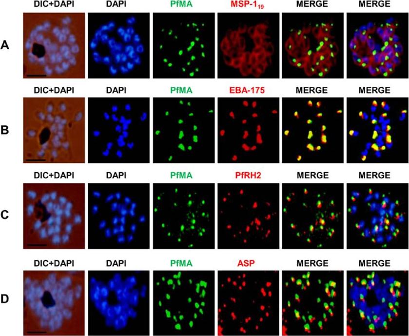
PfMA localization in schizont stages analyzed by fluorescence microscopy. Sub-cellular localization of PfMA was studied by co-immunostaining with surface protein (A), microneme (B), rhoptry (C, D) resident proteins. P. falciparum schizonts were co-immunostained with mouse anti-PfMA (green) and rabbit antibodies against one of the 4 marker proteins (EBA175/PfRH2, ASP, MSP-119) (red). The nuclei of schizont were stained with DAPI (blue) and slides were visualized by fluorescence microscope. All apical marker proteins and PfMA showed punctate staining in schizonts. PfMA was localized in the micronemes as it signal costained with micronemal marker EBA-175. The scale bar indicates 2 μm.Hans N, Singh S, Pandey AK, Reddy KS, Gaur D, Chauhan VS. Identification and Characterization of a Novel Plasmodium falciparum Adhesin Involved in Erythrocyte Invasion. PLoS One. 2013 8(9):e74790.
See original on MMP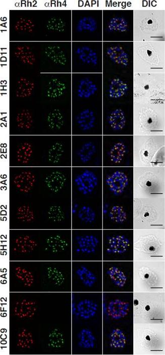
Localization of native PfRh4 was assessed by wide-field immunofluorescence assay using the anti-PfRh4 mAbs. PfRh4 (green) was co-stained with rhoptry protein, PfRh2 (red) and nuclei-stain DAPI (blue). DIC shows the differential interference contrast view of the same field. Scale bar = 5 μm. PfRh4, together with the other PfRh family of proteins, localize to the rhoptries of P. falciparum parasites in an apical localization that predominantly co-localized with PfRh2 another rhoptry protein.Lim NT, Harder MJ, Kennedy AT, Lin CS, Weir C, Cowman AF, Call MJ, Schmidt CQ, Tham WH. Characterization of Inhibitors and Monoclonal Antibodies that Modulate the Interaction between Plasmodium falciparum Adhesin PfRh4 with its Erythrocyte Receptor Complement Receptor 1. J Biol Chem. 2015 Aug 31. [Epub ahead of print]
See original on MMP
Specific amino acid residues in the cytoplasmic domain are essential for PfRh4 function. Localization of PfRh4 and PfRh2 in Rh4-WT tail, RH4-AMA1 tail (PfRh4 tail was replaced with the cytoplasmic sequence of AMA1), Rh4-mut4tail ) contains the mutations S1667A, S1674A, Y1680A and Y1684A) and RH4-mut5tail (contains similar mutations with an addition mutation at S1652A within the PfRh4 cytoplasmic tail) lines as detected using anti-PfRh4 monoclonal and anti-PfRh2 polyclonal antibodies. Parasite nuclei were stained with DAPI. Rh4-mut4 tail and Rh4-mut5 tail expressed PfRh4 and all showed an apical localization similar to rhoptry protein PfRh2Tham WH, Lim NT, Weiss GE, Lopaticki S, Ansell BR, Bird M, Lucet I, Dorin-Semblat D, Doerig C, Gilson PR, Crabb BS, Cowman AF. Plasmodium falciparum Adhesins Play an Essential Role in Signalling and Activation of Invasion into Human Erythrocytes. PLoS Pathog. 2015 Dec 22;11(12):e1005343.
See original on MMP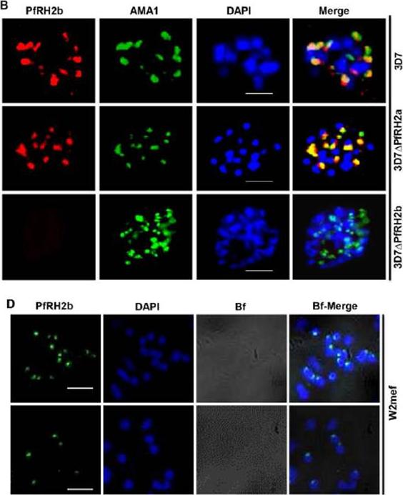
(B) IFA indicates the specificity of mAb A7 in schizont stage parasites. PfRH2b (PfRH2b, red) co-stained with AMA1 (AMA1, green), DAPI for nuclei (blue) in 3D7, 3D7ΔPfRH2a and 3D7ΔPfRH2b schizont. In the merged images, areas of overlap between the red and the green signals are shown in yellow. Scale bar indicated in white represents 2 μm. (D) Nonpermeabilized W2mef merozoites show surface expression of PfRH2b using mAb A7. PfRH2b shown green (RH2b A7), DAPI for nuclei (blue) and bright field (Bf) images shown in gray. Scale bar indicates 2 μm.Aniweh Y, Gao X, Gunalan K, Preiser PR. PfRH2b Specific Monoclonal Antibodies Inhibit Merozoite Invasion. Mol Microbiol. 2016 Jul 20.
See original on MMP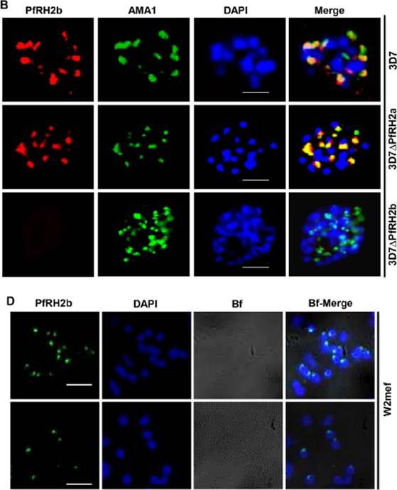
B. IFA indicates the specificity of mAb A7 in schizont stage parasites. PfRH2b (PfRH2b, red) co-stained with AMA1 (AMA1, green), DAPI for nuclei (blue) in 3D7, 3D7DPfRH2a and 3D7DPfRH2b schizont. In the merged images, areas of overlap between the red and the green signals are shown in yellow. Scale bar indicated in white represents 2 mm. D. Non-permeabilized W2mef merozoites show surface expression of PfRH2b using mAb A7. PfRH2b shown green (RH2b A7), DAPI for nuclei (blue) and bright field (Bf) images shown in gray. Scale bar indicates 2 mm. (3D7DPfRH2a (clone in which PfRH2a has been deleted), 3D7DPfRH2b (clone in which the PfRH2b gene has been deleted) parasites. The punctate staining pattern observed for A7 was in close proximity to the staining observed with an antibody targeting the micronemal protein Apical membrane antigen 1 (AMA1), indicating an apical location of PfRH2b within the merozoite.Aniweh Y, Gao X, Gunalan K, Preiser PR. PfRH2b specific monoclonal antibodies inhibit merozoite invasion. Mol Microbiol. 2016 Nov;102(3):386-404.
See original on MMPMore information
| PlasmoDB | PF3D7_1335400 |
| GeneDB | PF3D7_1335400 |
| Malaria Metabolic Pathways | Localisation images Pathways mapped to |
| Previous ID(s) | PF13_0198 |
| Orthologs | |
| Google Scholar | Search for all mentions of this gene |