PF3D7_1334500 MSP7-like protein (MSRP6)
Disruptability [+]
| Species | Disruptability | Reference | Submitter |
|---|---|---|---|
| P. falciparum 3D7 |
Possible |
20472690 | Theo Sanderson, Wellcome Trust Sanger Institute |
| P. falciparum 3D7 |
Refractory |
USF piggyBac screen (Insert. mut.) | USF PiggyBac Screen |
Mutant phenotypes [+]
| Species | Stage | Phenotype | Reference | Submitter |
|---|---|---|---|---|
| P. falciparum 3D7 | Asexual |
No difference |
20472690 | Theo Sanderson, Wellcome Trust Sanger Institute |
Imaging data (from Malaria Metabolic Pathways)
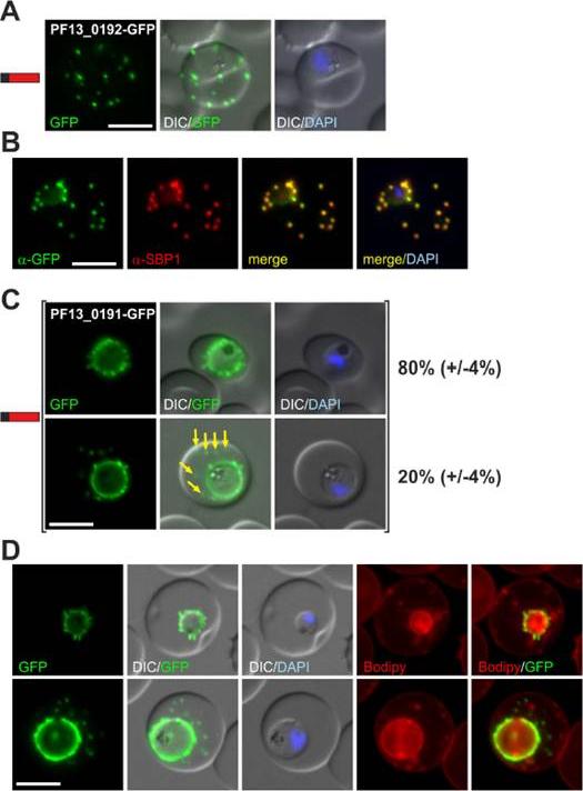
(A) Fluorescence pattern of PF13_0192-GFP. (B) Co-localisation IFA of PF13_0192-GFP with SBP1. (C) Fluorescence pattern of PF13_0191-GFP. Two panels are shown to demonstrate cells with (yellow arrows) and without additional foci of fluorescence in the host cell (ratio indicated in %, at least 50 cells were analysed on 3 occasions, standard deviation in brackets). (D) Bodipy-TR-C5-ceramide (Bodipy) stained parasites expressing PF13_0191-GFP. Protein structure in A and C indicated as in Figure 1. Nuclei were stained with DAPI. Size bars: 5 mm. PF13_0192-GFP was exported to foci in the host cell (A) that were confirmed to be Maurer’s clefts (B). PF13_0191-GFP localized to foci and mobile protrusions at the parasite periphery and in 20% (+/24%) of cells was also found in usually multiple mobile foci in the host cell with no apparent contact to the parasite periphery.Heiber A, Kruse F, Pick C, Grüring C, Flemming S, Oberli A, Schoeler H, Retzlaff S, Mesén-Ramírez P, Hiss JA, Kadekoppala M, Hecht L, Holder AA, Gilberger TW, Spielmann T. Identification of New PNEPs Indicates a Substantial Non-PEXEL Exportome and Underpins Common Features in Plasmodium falciparum Protein Export. PLoS Pathog. 2013 9(8):e1003546.
See original on MMP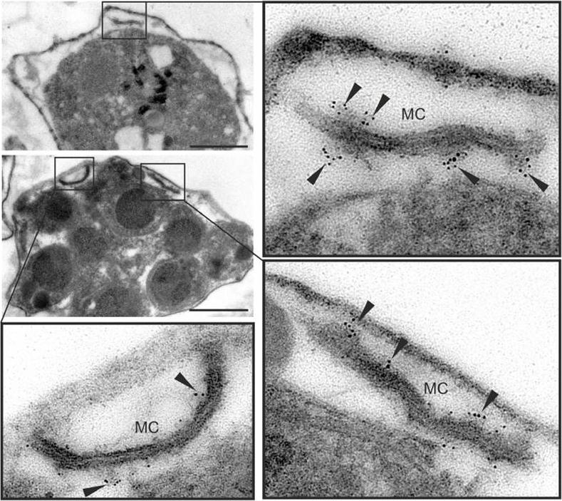
MSRP6 is a Maurer’s clefts protein expressed in trophozoites and schizonts. Pre-embedding immuno-EM of infected RBC, where the host cell cytosol has been released with tetanolysin, reacted with anti-MSRP6 antisera. Small panels show overviews with boxes highlighting individual Maurer’s clefts for which enlargements are shown. Gold label is indicated with arrowheads. Size bars: 1 mm. MSRP6 was detected in ‘cloudy’ structures at the outside of Maurer’s clefts.Heiber A, Kruse F, Pick C, Grüring C, Flemming S, Oberli A, Schoeler H, Retzlaff S, Mesén-Ramírez P, Hiss JA, Kadekoppala M, Hecht L, Holder AA, Gilberger TW, Spielmann T. Identification of New PNEPs Indicates a Substantial Non-PEXEL Exportome and Underpins Common Features in Plasmodium falciparum Protein Export. PLoS Pathog. 2013 9(8):e1003546
See original on MMP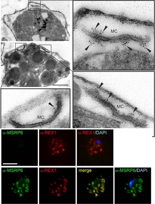
MSRP6 is a Maurer’s clefts protein expressed in trophozoites and schizonts. Pre-embedding immuno-EM of infected RBC, where the host cell cytosol has been released with tetanolysin, reacted with anti-MSRP6 antisera. Small panels show overviews with boxes highlighting individual Maurer’s clefts for which enlargements are shown. Gold label is indicated with arrowheads. Size bars: 1 mm. MSRP6 was detected in ‘cloudy’ structures at the outside of Maurer’s clefts. Lower panel: Co-localisation IFA using anti-REX1 and anti-MSRP6 antibodies with MSRP6 knock out parasites (top) and parental 3D7 parasites (bottom). Size bar: 5 mm. MSRP6 antiserum recognized foci in infected host cells representing Maurer’s clefts.Heiber A, Kruse F, Pick C, Grüring C, Flemming S, Oberli A, Schoeler H, Retzlaff S, Mesén-Ramírez P, Hiss JA, Kadekoppala M, Hecht L, Holder AA, Gilberger TW, Spielmann T. Identification of New PNEPs Indicates a Substantial Non-PEXEL Exportome and Underpins Common Features in Plasmodium falciparum Protein Export. PLoS Pathog. 2013 9(8):e1003546
See original on MMP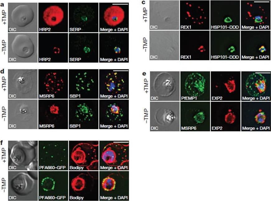
HSP101 is required for export of PEXEL and PNEP proteins. a, c, Immunofluorescence assay (IFA) of ring-stage 13F10 parasites with orwithout TMP (DDD-stabilizing small molecule trimethoprim). TMP was removed in late schizont stage and parasites were allowed to reinvade and grow 18–24 h before fixation with paraformaldehyde (a) or acetone (c). a, IFA of the exported PEXEL-containing protein HRP2. c, IFAs of the PNEP REX1, which colocalizes with HSP101DDD at the PVM in the absence of TMP. d, e, IFA of trophozoite-stage 13F10 parasites with or withoutTMP. TMP was removed in late ring stage and parasites were allowed to develop 12–24 h before fixation with acetone. f, Live fluorescence imaging of 13F10 parasites expressing a PFA660–GFP fusion and labelled with Bodipy TR Ceramide to demarcate the PVM (other membranes are also labelled). TMP treatment as in d, e. All scale bars, 5 mm. Images in c–f are representative of two independent experiments.Beck JR, Muralidharan V, Oksman A, Goldberg DE. PTEX component HSP101 mediates export of diverse malaria effectors into host erythrocytes. Nature. 2014 Jul 16. [Epub ahead of print]
See original on MMP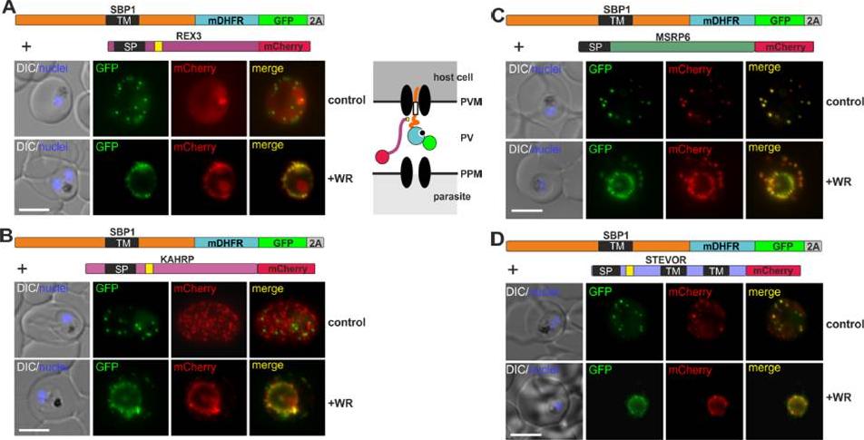
Different types of proteins pass through the same translocon. (A-D) Representative images of live P. falciparum parasites grown in the presence (+WR) or absence of WR (control), expressing the constructs shown schematically above each panel. The skip peptide is indicated by a grey box labelled 2A. The two proteins expressed from the same open reading frame are shown skipped. DIC, differential interference contrast. Size bars: 5 μm. A schematic of the co-block is shown for A. The yellow box indicates the mature PEXEL.Mesén-Ramírez P, Reinsch F, Blancke Soares A, Bergmann B, Ullrich AK, Tenzer S, Spielmann T. Stable Translocation Intermediates Jam Global Protein Export in Plasmodium falciparum Parasites and Link the PTEX Component EXP2 with Translocation Activity. PLoS Pathog. 2016 May 11;12(5):e1005618.
See original on MMPMore information
| PlasmoDB | PF3D7_1334500 |
| GeneDB | PF3D7_1334500 |
| Malaria Metabolic Pathways | Localisation images Pathways mapped to |
| Previous ID(s) | PF13_0192 |
| Orthologs | |
| Google Scholar | Search for all mentions of this gene |