PF3D7_1327600 nicotinate-nucleotide adenylyltransferase (NMNAT)
Disruptability [+]
| Species | Disruptability | Reference | Submitter | |
|---|---|---|---|---|
| P. falciparum 3D7 |
Possible |
USF piggyBac screen (Insert. mut.) | USF PiggyBac Screen | |
| P. berghei ANKA |
Refractory |
RMgm-512 | Imported from RMgmDB | |
Mutant phenotypes [+]
| Species | Stage | Phenotype | Reference | Submitter |
|---|---|---|---|---|
| P. berghei ANKA | Asexual |
Attenuated |
PlasmoGEM (Barseq) | PlasmoGEM |
Imaging data (from Malaria Metabolic Pathways)
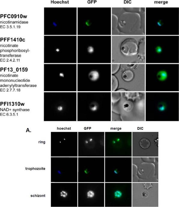
Live imaging of episomally expressed GFP tagged NAD+ metabolic enzymes are shown (GFP-fusion proteins are shown in green). Enzyme Commission numbers are provided for each enzyme. All images are of trophozoite stage parasites. Hoechst dye (shown in blue) was used to visualize the parasite nucleus. NAD+ pathway enzymes are primarily localized in the cytoplasm except for the parasite nicotinamidase, which localizes to the cytoplasm but concentrates mainly in the nucleus.Live imaging of the episomally expressed PfNico-GFP fusion. The nucleus is visualized with Hoechst staining. O'Hara JK, Kerwin LJ, Cobbold SA, Tai J, Bedell TA, Reider PJ, Llinás M. Targeting NAD+ Metabolism in the Human Malaria Parasite Plasmodium falciparum. PLoS One. 2014 Apr 18;9(4):e94061.
See original on MMP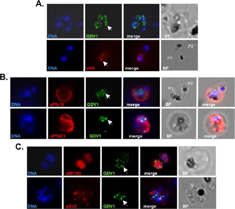
Subcellular localization of PfGDV1. Parasites transformed with GFP- or HA-tagged PfGDV1 were stained with DAPI (DNA stain) and the indicated anti-sera, and then examined by fluorescence microscopy. Images are shown of the DAPI stain (DNA), GFP-tagged PfGDV1 epifluorescence (GDV1), and antibodies specific for HA (aHA), Pfs16 (aPfs16), PfGE3 (aPfGE3), PfMCM2 (aMCM2), and PfSir2 (aSir2). The corresponding merged and bright field (BF) images are included on the right. PfGDV1 expression is indicated with an arrow; locations of parasites in the BF image are indicated with a P for parasite or S for schizont. A) A schizont (S) (Upper) expressing GDV1 and a doubly infected erythrocyte (Lower) with one parasite (P1) expressing HA-tagged PfGDV1 (aHA) and another negative (P2) for anti-HA antibodies. B) Costaining of parasites expressing GDV1 with early gameto-cytogenesis markers. A doubly infected erythrocyte (Upper) with one parasite (P1) positive for GDV1 and aPfs16 and the other (P2) negative for both. An erythrocyte (Lower) infected with a parasite (P) positive for GDV1 and aPfGE3. C) Colocalization of PfGDV1 with nuclear proteins. A doubly infected erythrocyte (Upper) with one parasite in the plane of the image (P1) and the other below (P2). Both P1 and P2 are positive for GDV1 and aMCM2. A schizont (S) (Lower) expressing GDV1 stained with aSir2.Eksi S, Morahan BJ, Haile Y, Furuya T, Jiang H, Ali O, Xu H, Kiattibutr K, Suri A, Czesny B, Adeyemo A, Myers TG, Sattabongkot J, Su XZ, Williamson KC. Plasmodium falciparum gametocyte development 1 (Pfgdv1) and gametocytogenesis early gene identification and commitment to sexual development. PLoS Pathog. 2012;8(10):e1002964.
See original on MMPMore information
| PlasmoDB | PF3D7_1327600 |
| GeneDB | PF3D7_1327600 |
| Malaria Metabolic Pathways | Localisation images Pathways mapped to |
| Previous ID(s) | 2008.m00035, PF13_0159 |
| Orthologs | PBANKA_1342800 , PCHAS_1347400 , PKNH_1201000 , PVP01_1226800 , PVX_116500 , PY17X_1347500 |
| Google Scholar | Search for all mentions of this gene |