PF3D7_1323500 plasmepsin V (PMV)
Disruptability [+]
| Species | Disruptability | Reference | Submitter | |
|---|---|---|---|---|
| P. falciparum 3D7 |
Refractory |
24983235 | Theo Sanderson, Wellcome Trust Sanger Institute | |
| P. falciparum 3D7 |
Refractory |
USF piggyBac screen (Insert. mut.) | USF PiggyBac Screen | |
| P. berghei ANKA |
Refractory |
RMgm-881 | Imported from RMgmDB | |
| P. berghei ANKA |
Refractory |
PlasmoGEM (Barseq) | PlasmoGEM | |
Mutant phenotypes [+]
| Species | Stage | Phenotype | Reference | Submitter |
|---|---|---|---|---|
| P. falciparum 3D7 | Asexual |
Cell cycle arrest |
30517136 DiCre-based approach. |
Theo Sanderson, Google AI |
Imaging data (from Malaria Metabolic Pathways)
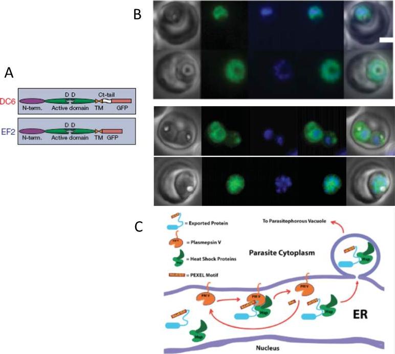
A. Schematic of PMV and C-terminal integrants. SP, signal peptide; N-term., N-terminal domain; Ct, C-terminal; TM, transmembrane region.B. Proteins destined for export are synthesized in the endoplasmic reticulum (ER) and cleaved at a conserved (PEXEL) motif, which allows translocation into the host cell via an ATP-driven translocon called the PTEX complex. We report that plasmepsin V, an ER aspartic protease recognizes the PEXEL motif and cleaves it at the correct site. Live fluorescence images of DC6 and EF2. Left to right: phase, GFP, DAPI, fluorescence merge and total merge. Scale bar, 2 mm.C. Schematic model of exported protein interactions. A PEXEL-containing protein is recognized by PMV in association with chaperones such as HSP 70 and HSP 101. PMV cleaves the PEXEL, releasing mature protein into the stewardship of the chaperones, which usher the protein through the secretory system to the translocon for export into the host erythrocyte.Russo I, Babbitt S, Muralidharan V, Butler T, Oksman A, Goldberg DE. Plasmepsin V licenses Plasmodium proteins for export into the host erythrocyte. Nature. 2010 463(7281):632-6.
See original on MMP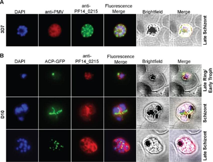
Cellular localization of HRD1. (A) HRD1 and plasmepsin V (PMV), an ER membrane marker, co-localize in P. falciparum. (B) At various stages of the parasite life cycle, HRD1 was co-stained with the nuclei (DAPI) and the apicoplast (ACP-GFP) in the P. falciparum strain D10. HRD1 was found within reticular structures outside the nuclear regions at the trophozoite and schizont stages of the parasite, similar to the physical attributes of the ER. In the late schizont stage HRD1 proteins reside within globular structures surrounding each budding merozoite in a pattern typical of the ER. These observations support that HRD1-E3 ubiquitin ligase resides in the ER membrane, consistent with a putative role in the ERAD pathway.Chung DW, Ponts N, Prudhomme J, Rodrigues EM, Le Roch KG. Characterization of the Ubiquitylating Components of the Human Malaria Parasite's Protein Degradation Pathway. PLoS One. 2012;7(8):e43477.
See original on MMP
Localization of plasmepsin V (PM V). Indirect immunofluorescence assays of PM V (green) in trophozoites (A) and a ~6N schizont (B) in formaldehyde-fixed blood smears. Nuclei were visualized with DAPI (pseudocolored red). (C, D) Colocalization of PM V and the ER protein BiP (C) or the Golgi marker ERD2 (D) in trophozoites. Bar, 2 mm. In trophozoites and schizonts, PM V was concentrated around the nucleus with lower levels dispersed in the surrounding cytoplasm (Fig. 5A and B). These patterns of fluorescence suggest that PM V resides in the endoplasmic reticulum (ER), which in P. falciparum has been shown to include the nuclear envelope.Klemba M, Goldberg DE. Characterization of plasmepsin V, a membrane-bound aspartic protease homolog in the endoplasmic reticulum of Plasmodium falciparum. Mol Biochem Parasitol. 2005 143:183-191. Copyright Elsevier 2010.
See original on MMP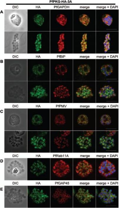
Subcellular location of PfPKG in mature schizonts. Dual immunofluorescent detection of PfPKG-HA in fixed smears of early and late schizonts of the PfPKG-HA-3A clone together with (A) GAPDH; (B) BiP; (C) PMV; (D) Rab11A and (E) GAP45. Representative images are shown for each antibody, together with bright field images (first column) and parasite nuclei stained with DAPI (in the merged image). Bars ,5 mm. Significant overlap between the PKG-HA and GAPDH in early and late blood stage schizonts (A), both are in the cytosol. PfPKG-HA also partially overlapped with that of the ER membrane protein PMV and that of the ER lumen marker PfBiP (B and C) in both early and late schizonts. Rab11A is thought to be involved in vesicle trafficking and localises to the rhoptries and the inner membrane complex (IMC) at the apical end of the merozoite. In mature schizonts, the location of PKG-HA appeared to be largely distinct from that of Rab11A (D). PKG-HA appeared to be absent from the IMC in segmented schizonts, since it did not colocalise with the IMC marker, the glideosome associated protein 45. Hopp CS, Flueck C, Solyakov L, Tobin A, Baker DA. Spatiotemporal and Functional Characterisation of the Plasmodium falciparum cGMP-Dependent Protein Kinase. PLoS One. 2012;7(11):e48206.
See original on MMP
Localization of EEA1WT-mCherry as a marker for PI(3)P (phosphatidylinositol-3-phosphate) in the parasite ER. EEA1 = early endosomal antigen 1. Transgenic parasites transfected with EEA1 were fixed, permeabilized and probed with anti-mCherry as well as antibodies to endogenous P. falciparum markers (green) of the ER, like BIP (top row) and plasmepsin V (Pl. V, second row); a marker of the Golgi (ERD2, third row) or the PVM (Exp-2, bottom row). EEA1WT (red) is seen in punctuate ‘spots’ within the ER. Dotted circles show location of red cell membrane. Dotted squares show regions magnified in the right panels. Parasite nuclei were stained with Hoechst 33342 (blue). Scale bars, 5 mm. The overlap between markers and EEA1 indicates the presence of PI(3)P in the respective regions.Bhattacharjee S, Stahelin RV, Speicher KD, Speicher DW, Haldar K. Endoplasmic Reticulum PI(3)P Lipid Binding Targets Malaria Proteins to the Host Cell. Cell. 2012 148(1-2):201-12.
See original on MMP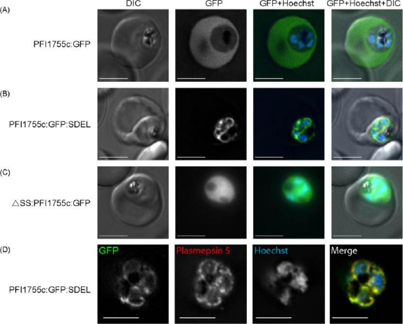
Localization of GFP chimeras in transfected parasites. (A) Localization of PFI1755c:GFP. GFP fluorescence, in live cells, is seen in the erythrocyte cytoplasm indicating that the protein is efficiently exported. (B) Localization of PFI1755c:GFP:SDEL. GFP fluorescence, in live cells, remains within the parasite and shows a characteristic ER distribution. (C) Localization of DSS:PFI1755c:GFP. GFP fluorescence, in live cells, is localized diffusely throughout the cytoplasm of the parasite. Panels show a DIC image, GFP fluorescence, GFP (green) and Hoechst (blue) fluorescence overlaid, and a merge of DIC, GFP (green) and Hoechst (blue) images, as indicated. The scale bar is 4mm. (D) Colocalization of PFI1755c:GFP:SDEL with the ER marker, plasmepsin 5. The GFP fluorescence in PFI1755c:GFP:SDEL expressing parasites was directly visualized and the localization of plasmepsin 5 was determined by indirect immunofluorescence, using an antibody raised against plasmepsin 5 . DNA was stained with Hoechst. In the merged image, GFP fluorescence is in green, plasmepsin 5 staining in red, and regions of co-localization in yellow. The scale bar is 4mm.Osborne AR, Speicher KD, Tamez PA, Bhattacharjee S, Speicher DW, Haldar K. The host targeting motif in exported Plasmodium proteins is cleaved in the parasite endoplasmic reticulum. Mol Biochem Parasitol. 2010 171:25-31. Copyright Elsevier 2010
See original on MMP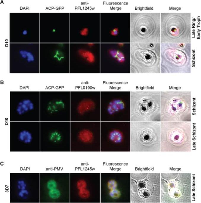
Localization studies of UBA (E1) and UBC (E2). (A,B) IFA experiments, using different parasite stages, show that PfUBA1 and PfUBC localize mainly to the cytosol. (C) When co-immunostained with plasmepsin V (PMV), a Plasmodium ER membrane protein marker, there was noticeable overlap between the two proteins, suggesting UBA1 recruitment to the ER as well. Both UBC and UBA1 are mainly dispersed throughout the cytoplasm with small aggregations scattered throughout the parasite. UBA1 was co-immuno-stained with PMV, an ER membrane marker, showing noticeable overlap between the two proteins.Chung DW, Ponts N, Prudhomme J, Rodrigues EM, Le Roch KG. Characterization of the Ubiquitylating Components of the Human Malaria Parasite's Protein Degradation Pathway. PLoS One. 2012;7(8):e43477.
See original on MMP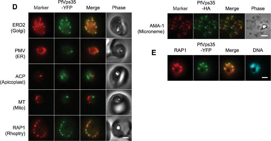
(D) Co-localization of PfVps35-YFP or PfVps35-HA and organellar markers in fixed parasites (except for MitoTracker, which was imaged live). PMV, plasmepsin V; ACP, acyl carrier protein; MT, MitoTracker Red CM-H2Xros; RAP1, rhoptry associated protein 1; AMA1, apical membrane antigen 1. The AMA1 panel shows free merozoites; all others are intraerythrocytic. Organelles labeled by the markers are indicated in parenthesis. Marker-derived fluorescence is pseudocolored red. (E) PfVps35-YFP is adjacent to developing rhoptries in a 2N parasite. Hoechst 33342 fluorescence (DNA) is pseudocolored cyan. In all panels, YFP fluorescence is pseudocolored green. Scale bars, 2 mm.Krai P, Dalal S, Klemba M. Evidence for a Golgi-to-Endosome Protein Sorting Pathway in Plasmodium falciparum. PLoS One. 2014 9(2):e89771.
See original on MMP
(D, Top) Immunofluorescence micrographs show rabbit a-PfPMV antibodies (Ra-PfPMV, green) label PfPMV in the ER. Colocalizations were performed with mouse a-PfPMV antibodies (Ma-PfPMV, red), shown previously to label PMV in the ER . (Middle) a-HA antibodies (red) label PfPMVHA (Top) and PvPMVHA (Bottom). P. vivax) in the parasite ER. (Bottom) a-HA antibodies (red) label PvPMVHA in the ER, as shown by clocalization with ERC (green). A strong perinuclear signal was observed in parasites expressing PvPMVHA or PfPMVHA (middle panels, red). Both proteins colocalized with rabbit anti-PfPMV antibodies, indicating the location was the ER. PvPMVHA also colocalized with ERC (bottom), as shown previously for PfPMVHA.Sleebs BE, Lopaticki S, Marapana DS, O'Neill MT, Rajasekaran P, Gazdik M, Günther S, Whitehead LW, Lowes KN, Barfod L, Hviid L, Shaw PJ, Hodder AN, Smith BJ, Cowman AF, Boddey JA. Inhibition of Plasmepsin V Activity Demonstrates Its Essential Role in Protein Export, PfEMP1 Display, and Survival of Malaria Parasites. PLoS Biol. 2014 12(7):e1001897.
See original on MMP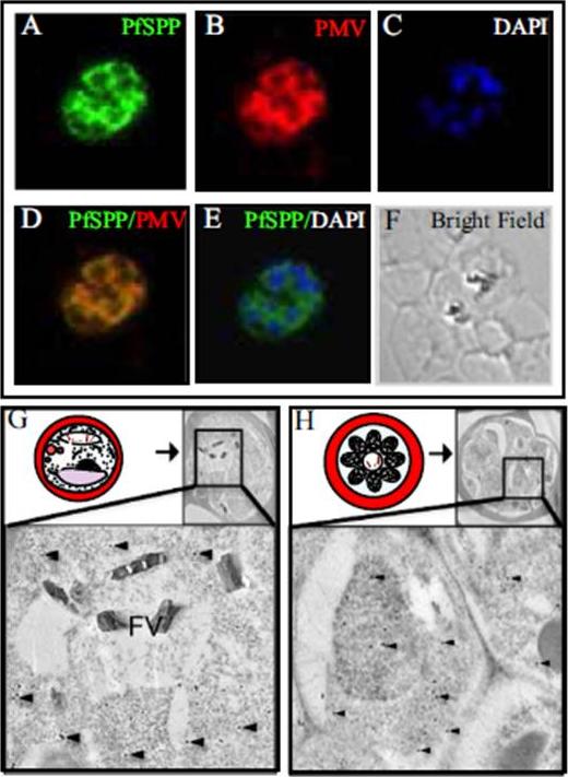
Co-localization of PfSPP (signal peptide peptidase) and Plasmepsin V in the endoplasmic reticulum. Indirect immunofluorescence microscopy using specific antibodies demonstrates PfSPP (green) co-localizes with a known parasite ER marker Plasmepsin V (red) in the Plasmodium falciparum (3D7) trophozoite. (A) Staining of PfSPP. (B) Staining of known ER marker, Plasmepsin V. (C) DAPI stain. (D) PfSPP and PMV overlay. (E) Overlay of PfSPP and DAPI stain. (F) Bright field microscopy. (G and H) Immunogold electron microscopy of PfSPP in the parasite infected erythrocytes. PfSPP is indicated by black dots and several spots are identified by black arrowheads. (G) Schematic illustration of a mature trophozoite stage parasite in the infected. PfSPP staining in the trophozoite shows a diffuse staining pattern throughout the perinuclear endomembrane system/ER. (H) Illustration of a late schizont-stage parasite in an infected erythrocyte. PfSPP staining in the late-schizont stage of parasite development shows staining throughout the perinuclear endomembrane system/ER. Baldwin M, Russo C, Li X, Chishti AH. Plasmodium falciparum signal peptide peptidase cleaves malaria heat shock protein 101 (HSP101). Implications for gametocytogenesis. Biochem Biophys Res Commun. 2014 Jul 10 [Epub ahead of print]
See original on MMP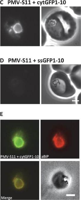
Using split GFP to establish the topology of Plasmepsin V. (C-D) Fluorescence (left) and phase contrast (right) images of P. falciparum parasites expressing PMV-S11 with cytGFP1-10 (C, lacks any targeting information) and PMV-S11 with ssGFP1-10 (D, fused to the signal sequence of BiP). Scale bar, 2 m. (E) Immunofluorescence colocalisation of the ER marker BiP (red) with the GFP fluorescence (green) in parasites expressing PMV-S11 with cytGFP 1–10. Scale bar, 2 m. Fluorescent parasites were observed when PMV-S11 was co-expressed with cytGFP1-10 (C), but not when co-expressed with ssGFP1-10 (D). ER localisation of the PMV-S11- cytGFP1-10 complex was confirmed by immunofluorescence analysis; the GFP signal in parasites expressing PMV-S11 with cytGFP1-10 showed extensive colocalisation with the ER marker BiP.Tarr SJ, Osborne AR. Experimental determination of the membrane topology of the Plasmodium protease plasmepsin v. PLoS One. 2015 7;10(4):e0121786.
See original on MMP
Sub-cellular distribution and localization of PfHsp60 (continued). (A) IFA of localization PfHsp60 in P. falciparum infected erythrocytes with (i) Propidium iodide (Nuclear marker), (ii) Mitotracker, (iii) PfCytochrome C (Mitochondrial marker), (iv) PfUROD (Apicoplast marker), (v) PfPlasmepsin V (PfPMV, endoplasmic reticulum marker), and (vi) PfHsp70-1 (Cytosol marker). PfHsp60 is stained with FITC-conjugated secondary antibody in all parts, while PfUROD, PfPlasmepsin V, PfCytochrome C, and PfHsp70-1 are stained with TRITC-cojugated secondary antibody in (iii), (iv), and (v). In all images, white arrow indicates the erythrocyte membrane. Part (i) shows that PfHsp60 is present only in the extra-nuclear compartment. Parts (ii) and (iii) show that PfHsp60 is present in the cytoplasm as well as in the mitochondria. The yellow signal in the merged panel shows that there is some co-localization. Part (iv) shows a lack of signal overlap with PfUROD in the merge panel. Part (v) shows a lack of signal overlap with PfPlasmepsin V in the merge panel, which rules out its localization in endoplasmic reticulum. Part (vi) shows that there is co-localization between the cytosolic protein PfHsp70-1 and PfHsp60, which indicates that PfHsp60 indeed accumulates in the cytosolPadma Priya P, Grover M, Tatu US, Natarajan V. Characterization of Precursor PfHsp60 in Plasmodium falciparum Cytosol during Its Asexual Development in Human Erythrocytes. PLoS One. 2015 Aug 28;10(8):e0136401. PMID:
See original on MMP
(c) p40PX-GFP localizes to the cytoplasm and to membranes of the food vacuole (labelled with anti-CRT) and apicoplast (labelled with anti-ACP). STEVORss-p40PX-GFP localizes to the PV in P. falciparum-infected erythrocytes, as shown by co-localization with anti-EXP2, and not in the ER, which was labelled with anti-PMV. If PI(3)P was located within the ER, this chimera would be expected to remain within the ER via p40PX binding. Fusion to an ER-retention signal (STEVORss-p40PX-GFPSDEL) retains the protein in ER, co-localization with anti-ERC. Scale bar, 5 mm.(d) Localization of Hrs-GFP to the cytoplasm and membranes of food vacuole (labelled anti-CRT) and apicoplast (labelled anti-ACP). STEVORss-Hrs-GFP localizes to the ER as shown by co-localization with anti-ERC; however, mutation of residues necessary for lipid binding in both Hrs FYVE fingers (STEVORss-HrsC215S-GFP) does not abrogate ER localization, demonstrating PI(3)P-independent retention in ER. Addition of ER retention signal (STEVORss-Hrs-GFPSDEL) also causes ER localization. Scale bar, 5 mm. Boddey JA, O'Neill MT, Lopaticki S, Carvalho TG, Hodder AN, Nebl T, Wawra S,van West P, Ebrahimzadeh Z, Richard D, Flemming S, Spielmann T, Przyborski J,Babon JJ, Cowman AF. Export of malaria proteins requires co-translationalprocessing of the PEXEL motif independent of phosphatidylinositol-3-phosphatebinding. Nat Commun. 2016 Feb 1;7:10470.
See original on MMPMore information
| PlasmoDB | PF3D7_1323500 |
| GeneDB | PF3D7_1323500 |
| Malaria Metabolic Pathways | Localisation images Pathways mapped to |
| Previous ID(s) | PF13_0133 |
| Orthologs | PBANKA_1338700 , PCHAS_1343300 , PKNH_1205300 , PVP01_1231100 , PVX_116695 , PY17X_1343400 |
| Google Scholar | Search for all mentions of this gene |