PF3D7_1320600 ras-related protein Rab-11A (RAB11a)
Disruptability [+]
| Species | Disruptability | Reference | Submitter | |
|---|---|---|---|---|
| P. falciparum 3D7 |
Refractory |
USF piggyBac screen (Insert. mut.) | USF PiggyBac Screen | |
| P. berghei ANKA |
Refractory |
RMgm-298 | Imported from RMgmDB | |
Mutant phenotypes [+]
None reported yet. Please press the '+' button above to add one.Imaging data (from Malaria Metabolic Pathways)
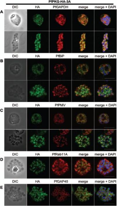
Subcellular location of PfPKG in mature schizonts. Dual immunofluorescent detection of PfPKG-HA in fixed smears of early and late schizonts of the PfPKG-HA-3A clone together with (A) GAPDH; (B) BiP; (C) PMV; (D) Rab11A and (E) GAP45. Representative images are shown for each antibody, together with bright field images (first column) and parasite nuclei stained with DAPI (in the merged image). Bars ,5 mm. Significant overlap between the PKG-HA and GAPDH in early and late blood stage schizonts (A), both are in the cytosol. PfPKG-HA also partially overlapped with that of the ER membrane protein PMV and that of the ER lumen marker PfBiP (B and C) in both early and late schizonts. Rab11A is thought to be involved in vesicle trafficking and localises to the rhoptries and the inner membrane complex (IMC) at the apical end of the merozoite. In mature schizonts, the location of PKG-HA appeared to be largely distinct from that of Rab11A (D). PKG-HA appeared to be absent from the IMC in segmented schizonts, since it did not colocalise with the IMC marker, the glideosome associated protein 45. Hopp CS, Flueck C, Solyakov L, Tobin A, Baker DA. Spatiotemporal and Functional Characterisation of the Plasmodium falciparum cGMP-Dependent Protein Kinase. PLoS One. 2012;7(11):e48206.
See original on MMP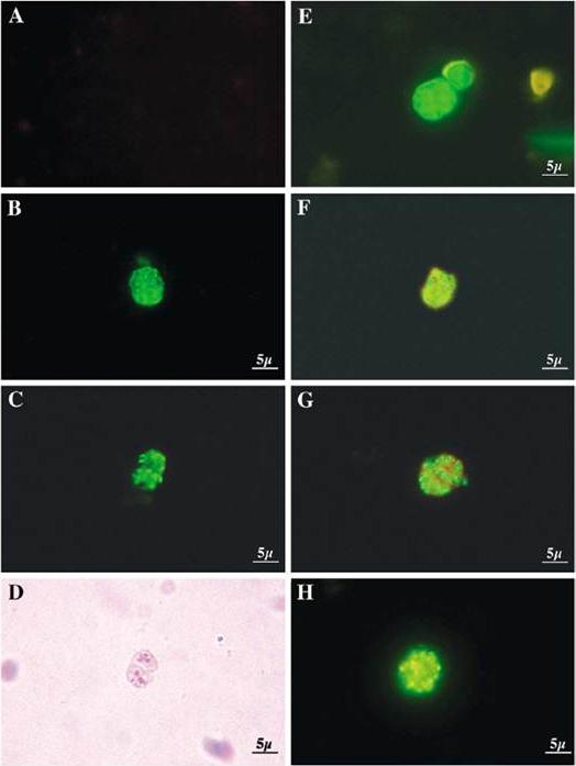
Different PfRabs have different sub-cellular distributions in schizont-infected erythrocytes. Panel (A) shows control rabbit pre-immune serum. Panel (B) shows a multinucleated schizont decorated with anti-PfRab1A antibodies giving a perinuclear staining. Panels (C) and (D) (phase contrast) show the pattern obtained with the anti-PfRab11A antibodies. Clearly, PfRab1A and PfRab11A display different distributions. Panel (E) shows the pattern obtained with anti-Pf39 (ERC) antibodies specific for the endoplasmic reticulum. Panel (F) shows a double-labelling with anti-PfRab1A (green) and Pf39 (red) antibodies, where colocalization stains yellow. Panel (G) shows a double-labelling with anti-PfRab11A (green) and anti-Pf39 (red) and no co-localization is observed. Panel (H) shows anti-PfRab6 (green) and anti-Pf39 (red), where co-localization stains yellow.Quevillon E, Spielmann T, Brahimi K, Chattopadhyay D, Yeramian E, Langsley G. The Plasmodium falciparum family of Rab GTPases. Gene. 2003 306:13-25.
See original on MMPMore information
| PlasmoDB | PF3D7_1320600 |
| GeneDB | PF3D7_1320600 |
| Malaria Metabolic Pathways | Localisation images Pathways mapped to |
| Previous ID(s) | PF13_0119 |
| Orthologs | PBANKA_1418900 , PCHAS_1420700 , PKNH_1421200 , PVP01_1421400 , PVX_122840 , PY17X_1420600 |
| Google Scholar | Search for all mentions of this gene |