PF3D7_1307500 conserved Plasmodium protein, unknown function
Disruptability [+]
| Species | Disruptability | Reference | Submitter |
|---|---|---|---|
| P. falciparum 3D7 |
Refractory |
USF piggyBac screen (Insert. mut.) | USF PiggyBac Screen |
Mutant phenotypes [+]
None reported yet. Please press the '+' button above to add one.Imaging data (from Malaria Metabolic Pathways)
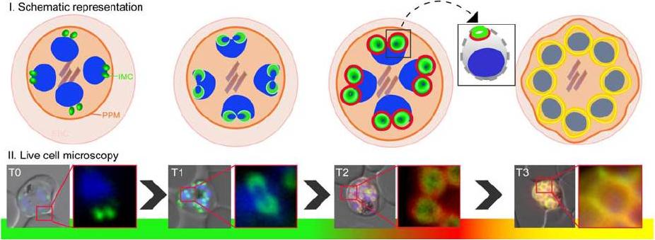
IMC biogenesis during blood stage proliferation in P. falciparum. Schematic representation and live cell microscopy of the IMC dynamics during merozoite development (T0-T3). Two IMC proteins with distinct phenotypes where either tagged with GFP (the group A (classical glideosome components) protein MAL13P1.130, green) or mCherry (the group B protein (proteins of the Alveolin family as well as MAL13P1.228; appear as rings with a wider diameter surrounding the already established compartment) PF13_0039, red). T0: nascent IMC compartments visible as two dots in young schizonts. T1: IMC forms cramp-like structures. T2: IMC is enlarged and group B protein (red) emerges at the proximal rim; square: highlighting the spatial arrangement of group A and B proteins. T3: The IMC represented by group A and B proteins fully surrounds the mature merozoite.Kono M, Prusty D, Parkinson J, Gilberger TW. The apicomplexan inner membrane complex. Front Biosci (Landmark Ed). 2013 Jun 1;18:982-92. Review.
See original on MMPMore information
| PlasmoDB | PF3D7_1307500 |
| GeneDB | PF3D7_1307500 |
| Malaria Metabolic Pathways | Localisation images Pathways mapped to |
| Previous ID(s) | PF13_0039 |
| Orthologs | PBANKA_1406000 , PCHAS_1407900 , PKNH_1407800 , PVP01_1408400 , PVX_122207 , PY17X_1407700 |
| Google Scholar | Search for all mentions of this gene |