PF3D7_1301700 Plasmodium exported protein (hyp8), unknown function (GEXP07)
Disruptability [+]
| Species | Disruptability | Reference | Submitter |
|---|---|---|---|
| P. falciparum 3D7 |
Refractory |
18614010 | Theo Sanderson, Wellcome Trust Sanger Institute |
| P. falciparum 3D7 |
Possible |
USF piggyBac screen (Insert. mut.) | USF PiggyBac Screen |
| P. falciparum 3D7 |
Possible |
https://www.biorxiv.org/content/10.1101/741033v1
Assisted by Cas9. "Disruption of VCAP1 causes Maurer’s cleft fragmentation, aberrant knobs, ablation of 38 PfEMP1 surface expression and loss of the PfEMP1 directed adhesion. VCAP1-disrupted parasite lines have 39 a growth advantage compared to wildtype parasites; and the infected RBCs are more deformable 40 and more osmotically fragile. " |
Theo Sanderson, Francis Crick Institute |
Mutant phenotypes [+]
| Species | Stage | Phenotype | Reference | Submitter |
|---|---|---|---|---|
| P. falciparum 3D7 | Asexual |
Altered cytoadherence |
https://www.biorxiv.org/content/10.1101/741033v1
Assisted by Cas9. "Disruption of VCAP1 causes Maurer’s cleft fragmentation, aberrant knobs, ablation of 38 PfEMP1 surface expression and loss of the PfEMP1 directed adhesion. VCAP1-disrupted parasite lines have 39 a growth advantage compared to wildtype parasites; and the infected RBCs are more deformable 40 and more osmotically fragile. " |
Theo Sanderson, Francis Crick Institute |
Imaging data (from Malaria Metabolic Pathways)
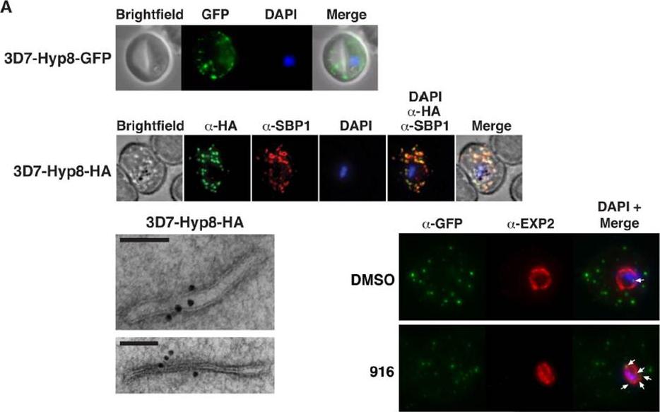
PMV inhibition impairs protein export, PfEMP1 display, and cytoadherence. (A) (Top) Immunofluorescent micrographs show Hyp8-GFP (exported protein hyp8),is exported and localizes within puncta in the infected erythrocyte. (Middle) Hyp8-HA localizes within SBP1-containing MCs. (Right) Immunoelectron microscopy shows Hyp8-HA localization at MCs. Scale bar is 100 nm. Immunofluorescence microscopy showed Hyp8-GFP is exported and colocalizes with SBP1 in MCs(B) Maximum intensity projection micrographs showing export of Hyp8-GFP to MCs and secretion of EXP2 to the parasitophorous vacuole membrane following treatment with DMSO or 916 (50 mM). Puncta of nonexported GFP within the parasite and vacuole is shown (arrows)Sleebs BE, Lopaticki S, Marapana DS, O'Neill MT, Rajasekaran P, Gazdik M, Günther S, Whitehead LW, Lowes KN, Barfod L, Hviid L, Shaw PJ, Hodder AN, Smith BJ, Cowman AF, Boddey JA. Inhibition of Plasmepsin V Activity Demonstrates Its Essential Role in Protein Export, PfEMP1 Display, and Survival of Malaria Parasites. PLoS Biol. 2014 12(7):e1001897.
See original on MMP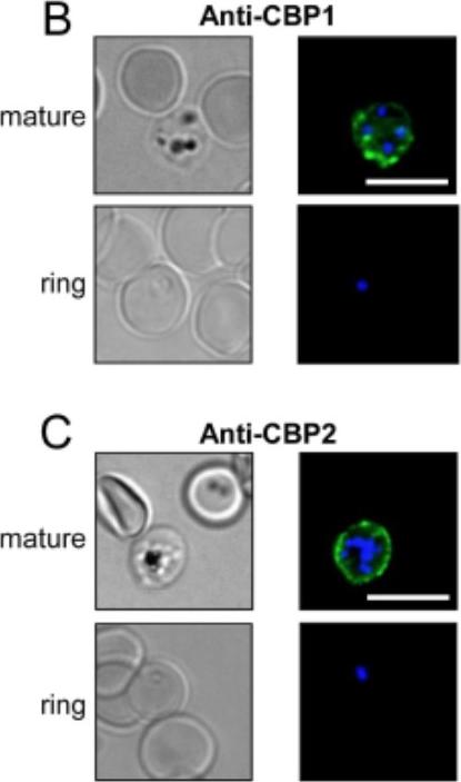
The antibodies raised against CBP1 and CBP2 stain the external membrane of RBC infected by mature 3D7 Plasmodium falciparum strain. (B,C) 3D7-iRBC were visualized by transmitted light, by staining with anti-CBP1 (B) or anti-CBP2 (C) antisera (green) and by Hoechst staining (blue) using either RBC infected by 3D7 strain at mature stages (80% parasitaemia, after gel flotation) or at early stage (4,5% parasitaemia, 4% ring). Bar = 10 μ m.Hermand P, Cicéron L, Pionneau C, Vaquero C, Combadière C, Deterre P. Plasmodium falciparum proteins involved in cytoadherence of infected erythrocytes to chemokine CX3CL1. Sci Rep. 2016 6:33786.
See original on MMPMore information
| PlasmoDB | PF3D7_1301700 |
| GeneDB | PF3D7_1301700 |
| Malaria Metabolic Pathways | Localisation images Pathways mapped to |
| Previous ID(s) | MAL13P1.61 |
| Orthologs | |
| Google Scholar | Search for all mentions of this gene |