PF3D7_1301400 Plasmodium exported protein (hyp12), unknown function (HYP12)
Disruptability [+]
| Species | Disruptability | Reference | Submitter |
|---|---|---|---|
| P. falciparum 3D7 |
Possible |
18614010 | Theo Sanderson, Wellcome Trust Sanger Institute |
| P. falciparum 3D7 |
Possible |
USF piggyBac screen (Insert. mut.) | USF PiggyBac Screen |
Mutant phenotypes [+]
None reported yet. Please press the '+' button above to add one.Imaging data (from Malaria Metabolic Pathways)
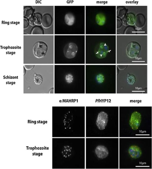
PfHYP12 is exported to the RBC cytoplasm and localizes to distinct intra-parasitic structures. Upper panel: Epifluorescence microscopy of erythrocytes infected with PfHYP12-GFP expressing 3D7 parasites. Images of live cells infected with parasites at three distinct stages are displayed. Merge image: Green, GFP; blue, Hoechst. DIC. Lower panel: Localization of PfHYP12-GFP by IFA. Cells were prepared for microscopy from asynchronous blood stage cultures. Representative co-immunofluorescence pictures are shown for two different stages of infection. Acetone/methanol (90%/10%) fixed smears were stained with mouse a-MAHRP1 (1:1000) (center left) and chicken a-GFP (1:500, Abcam) antibodies (center right) followedby incubation with Alexa Fluor-conjugated secondary antibodies in order to visualize PfHYP12-GFP and Maurer’s clefts, respectively. Merge image: red, MAHRP1; green, GFP; blue, Hoechst. Scale bars: 10 mm. Petersen W, Matuschewski K, Ingmundson A. Trafficking of the signature protein of intra-erythrocytic Plasmodium berghei-induced structures, IBIS1, to P. falciparum Maurer's clefts. Mol Biochem Parasitol. 2015 200(1-2):25-29. PMID:
See original on MMPMore information
| PlasmoDB | PF3D7_1301400 |
| GeneDB | PF3D7_1301400 |
| Malaria Metabolic Pathways | Localisation images Pathways mapped to |
| Previous ID(s) | PF13_0073 |
| Orthologs | |
| Google Scholar | Search for all mentions of this gene |