PF3D7_1254200 rifin (RIF)
Disruptability [+]
| Species | Disruptability | Reference | Submitter |
|---|---|---|---|
| P. falciparum 3D7 |
Refractory |
USF piggyBac screen (Insert. mut.) | USF PiggyBac Screen |
Mutant phenotypes [+]
None reported yet. Please press the '+' button above to add one.Imaging data (from Malaria Metabolic Pathways)
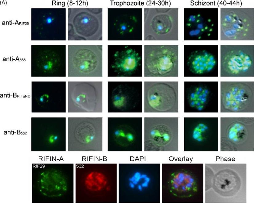
Indirect immunofluorescence assay in asexual stages of the FCR3S1.2 clone. A-type RIFINs identified by rabbit anti-A565 and rat anti-ARIF29, were found to be associated with the parasite and transported to the surface of infected erythrocytes via Maurer’s clefts. B-type RIFINs, identified by rabbit anti-B562 and mouse anti-BRIFDNC appeared to be mostly retained inside the parasite.Lower panel: A- and B-type RIFINs are both expressed in the same IE as detected by anti-ARIF29 (green) and anti-B562 (red) antisera, respectivelyPetter M, Haeggström M, Khattab A, Fernandez V, Klinkert MQ, Wahlgren M. Variant proteins of the Plasmodium falciparum RIFIN family show distinct subcellular localization and developmental expression patterns. Mol Biochem Parasitol. 2007 156:51-61. Copyright Elsevier 2009.
See original on MMP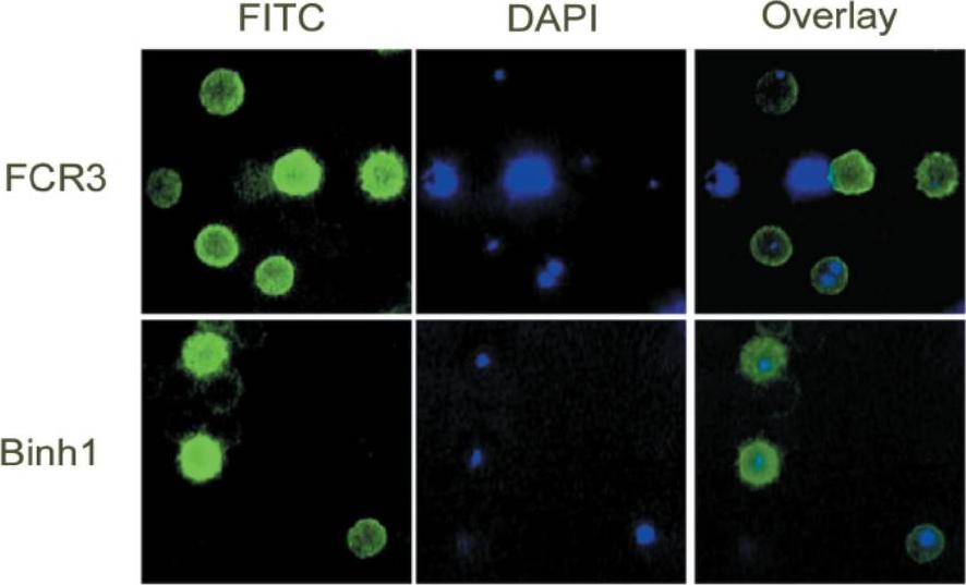
Indirect fluorescence assay of fixed IEs. (A) Laboratory strains FCR3 (top panels) and Binh1 (bottom panels) were stained with affinity-purified anti-rif-29 antibodies, and the secondary antibody was FITC-conjugated goat anti-human immunoglobulins (green; left panels). Parasite nuclei were visualized with DAPI (blue; middle panels), smaller spots are representative of the ring stages and larger spots are trophozoites. Both images were merged to show concordance (right panels). The protein was localized by fluorescence microscopy on the membrane of ring and young trophozoite-infected erythrocytesAbdel-Latif MS, Khattab A, Lindenthal C, Kremsner PG, Klinkert MQ. Recognition of variant Rifin antigens by human antibodies induced during natural Plasmodium falciparum infections. Infect Immun. 2002 70:7013-21. PMID:
See original on MMP
Upper panel: Indirect immunofluorescence assay in asexual stages of the FCR3S1.2 clone. A-type RIFINs (e.g., PFL2615w, PFD1240w), identified by rabbit anti-A565 and rat anti-ARIF29, were found to be associated with the parasite and transported to the surface of infected erythrocytes via Maurer’s clefts. B-type RIFINs (e.g., PFI0050c, PFE1630w), identified by rabbit anti-B562 and mouse anti-BRIFDNC appeared to be mostly retained inside the parasite.Lower panel: A- and B-type RIFINs are both expressed in the same IE as detected by anti-ARIF29 (green) and anti-B562 (red) antisera, respectivelyPetter M, Haeggström M, Khattab A, Fernandez V, Klinkert MQ, Wahlgren M. Variant proteins of the Plasmodium falciparum RIFIN family show distinct subcellular localization and developmental expression patterns. Mol Biochem Parasitol. 2007 156:51-61. Copyright Elsevier 2009
See original on MMPMore information
| PlasmoDB | PF3D7_1254200 |
| GeneDB | PF3D7_1254200 |
| Malaria Metabolic Pathways | Localisation images Pathways mapped to |
| Previous ID(s) | 2277.t00521, MAL12P1.518, PFL2615w |
| Orthologs | |
| Google Scholar | Search for all mentions of this gene |