PF3D7_1254100 stevor
Disruptability [+]
| Species | Disruptability | Reference | Submitter |
|---|---|---|---|
| P. falciparum 3D7 |
Possible |
USF piggyBac screen (Insert. mut.) | USF PiggyBac Screen |
Mutant phenotypes [+]
None reported yet. Please press the '+' button above to add one.Imaging data (from Malaria Metabolic Pathways)
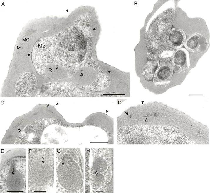
Immunoelectron microscopic localization of STEVORs. Ultrathin sections of schizont stage-IE were analyzed using anti-STEVOR (anti-PFL2610w) antibody. Gold labelling is observed on the IE surface (A and C, black arrowheads), merozoite surface (A, black arrows), in MC (A, C and D, white arrowheads), rhoptries (A, white arrows) and rhoptry neck (E-H, white arrows). No gold particles are observed with pre-immune serum, shown for an IE containing developing merozoites (B). Scale bars are 0.5 μm for A and 1 μm for B-H.Khattab A, Bonow I, Schreiber N, Petter M, Schmetz C, Klinkert MQ. Plasmodium falciparum variant STEVOR antigens are expressed in merozoites and possibly associated with erythrocyte invasion. Malar J. 2008 Jul 23;7:137.
See original on MMP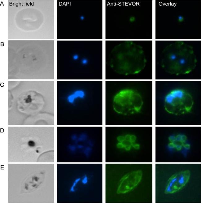
IFA images of methanol-fixed slides of the field isolate Gb337 in (A) early ring, (B) ring, (C) trophozoite, (D) schizont and (E) gametocyte stages visualized using anti-STEVOR (anti-PFL2610w) antibody. The localization of STEVOR proteins to the merozoite surface and the rhoptries together with its prevalence as a released component in the invading merozoite suggest a role of these antigens in adhesion and/or immune evasion in the erythrocyte invasion process.Khattab A, Bonow I, Schreiber N, Petter M, Schmetz C, Klinkert MQ. Plasmodium falciparum variant STEVOR antigens are expressed in merozoites and possibly associated with erythrocyte invasion. Malar J. 2008 Jul 23;7:137.
See original on MMP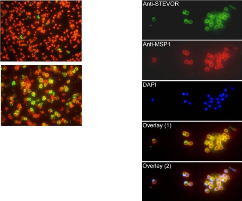
Left panel: Recognition of STEVORs in non-permeabilized and saponin-permeabilized schizont infected erythrocytes (IE). Fluorescence microscopy image of the schizont IE that were recognized by anti-PFL2610w antibody. Minimal binding of anti-PFL2610w antibody to the surface of schizont IE was observed. the anti-PFL2610w can only recognize STEVOR variants that are associated with internal schizont IE structures but not those presumably expressed on the surface. Right panel: Colocalization of STEVORs with MSP-1 at the free merozoite membranes. Fluorescence staining using anti-PFL2610w and anti-MSP-1 antibodies was analysed in free merozoites. (B) Alexa 488 stained STEVORs, (C) Alexa 594 stained MSP-1, (D) DAPI stained parasite nuclei, (E) the overlay of STEVORs and MSP-1 (overlay 1) and (F) the overlay of STEVORs, MSP-1 and nuclei (overlay 2) images are shown. Khattab A, Meri S. Exposure of the Plasmodium falciparum clonally variant STEVOR proteins on the merozoite surface. Malar J. 2011 Mar 14;10:58.
See original on MMP
b Co-localization of α-RIF44, STEVOR α-PFL2610w and α-PfMC-2TM-SC (green) with human spectrin (red). c Co-localization of α-RIF44, STEVOR α-PFC0025c and α-PfMC-2TM-CT (green) with SBP1 (red).Bachmann A, Scholz JA, Janßen M, Klinkert MQ, Tannich E, Bruchhaus I, Petter M. A comparative study of the localization and membrane topology of members of the RIFIN, STEVOR and PfMC-2TM protein families in Plasmodium falciparum-infected erythrocytes. Malar J. 2015 Jul 14:274.
See original on MMP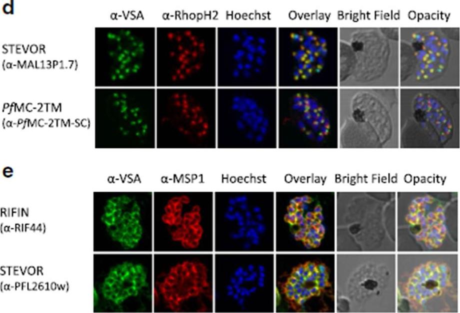
d Co-localization of STEVOR α-MAL13P1.7 or α-PfMC-2TM-SC (green) with the rhoptry marker RhopH2 (red). e Co-localization of α-RIF44 and STEVOR α-PFL2610w (green) with the merozoite surface protein MSP1 (red). Variant surface antigens (VSA).Bachmann A, Scholz JA, Janßen M, Klinkert MQ, Tannich E, Bruchhaus I, Petter M. A comparative study of the localization and membrane topology of members of the RIFIN, STEVOR and PfMC-2TM protein families in Plasmodium falciparum-infected erythrocytes. Malar J. 2015 Jul 14:274.
See original on MMP
Localization of small VSA in infected erythrocytes using confocal immunofluorescence analysis. a Asexual parasites of the 3D7 parasite clone at the trophozoite and schizont stages were fixed with methanol and small VSA localization was visualized using antibodies directed against RIFIN (α-RIF40.2, α-RIF44), STEVOR (α-PFL2610w, α-MAL13P1.7, α-PFC0025c, α-PFA0750w) and PfMC-2TM (α-PfMC-2TM-SC, α-PfMC-2TM-CT) proteins (green). Nuclei were stained with Hoechst33342 (blue).Bachmann A, Scholz JA, Janßen M, Klinkert MQ, Tannich E, Bruchhaus I, Petter M. A comparative study of the localization and membrane topology of members of the RIFIN, STEVOR and PfMC-2TM protein families in Plasmodium falciparum-infected erythrocytes. Malar J. 2015 Jul 14:274.
See original on MMPMore information
| PlasmoDB | PF3D7_1254100 |
| GeneDB | PF3D7_1254100 |
| Malaria Metabolic Pathways | Localisation images Pathways mapped to |
| Previous ID(s) | 2277.t00520, MAL12P1.517, PFL2610w |
| Orthologs | |
| Google Scholar | Search for all mentions of this gene |