PF3D7_1252100 rhoptry neck protein 3 (RON3)
Disruptability [+]
| Species | Disruptability | Reference | Submitter |
|---|---|---|---|
| P. falciparum 3D7 |
Refractory |
USF piggyBac screen (Insert. mut.) | USF PiggyBac Screen |
Mutant phenotypes [+]
| Species | Stage | Phenotype | Reference | Submitter |
|---|---|---|---|---|
| P. falciparum 3D7 | Asexual |
Cell cycle arrest |
31289187 'Exported proteins, such as ring-infected erythrocyte surface antigen (RESA), failed to be transported in parasites in which the parasite rhoptry protein RON3 was conditionally disrupted. RON3-deficient parasites also failed to develop beyond the ring stage, and glucose uptake was significantly decreased.' |
Theo Sanderson, Francis Crick Institute |
| P. falciparum 3D7 | Asexual |
Invasion defect |
37457963 (Knock down)
We demonstrated that RON3 is essential for blood-stage parasite survival, and knockdown of RON3 C-terminal fragment expression from the early schizont stage induces a defect in erythrocyte invasion and the subsequent development of ring stage parasites. |
Theo Sanderson, Francis Crick Institute |
Imaging data (from Malaria Metabolic Pathways)

Rhoptry body localization of PfRON3 by immunoelectron microscopy. Longitudinally sectioned merozoites in mature schizont were labeled with rabbit anti-PfRON3_2 antibodies followed by secondary antibody conjugated with gold particles. The image shows that the signals of gold particles were restricted to the rhoptry body of merozoite. Bars represent 500 nm.Ito D, Han ET, Takeo S, Thongkukiatkul A, Otsuki H, Torii M, Tsuboi T. Plasmodial ortholog of Toxoplasma gondii rhoptry neck protein 3 is localized to the rhoptry body. Parasitol Int. 2011 60(2):132-8.
See original on MMP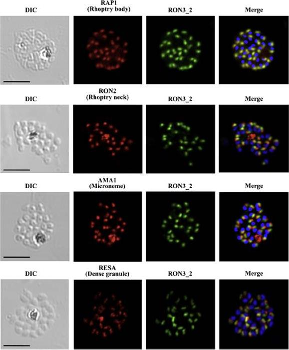
PfRON3 is expressed at the apical end of Plasmodium merozoites. Schizont and merozoite stage parasites were dual-labeled with antisera against PfRON3_2 and either PfRAP1 (rhoptry body marker), or PfRON2 (rhoptry neck marker), or PfAMA1 (microneme marker), or PfRESA (dense granule marker). Nuclei are visualized with DAPI in merged images shown in the right panels. Bars represent 5 μm. PfRON3 is localized to the but in the rhoptry body.Ito D, Han ET, Takeo S, Thongkukiatkul A, Otsuki H, Torii M, Tsuboi T. Plasmodial ortholog of Toxoplasma gondii rhoptry neck protein 3 is localized to the rhoptry body. Parasitol Int. 2011 60(2):132-8.
See original on MMP
PfRON3 exists in parasitophorous vacuole in ring stage parasites. (A) Ring stage parasites were dual-labeled with antisera against PfRON3_1 and either PfRAP1 (PV marker), PfEXP2 (PVM marker), PfRON2, or PfRON4. Nuclei are visualized with DAPI in merged images shown in the right panels. Bars represent 2.5 μm.Ito D, Han ET, Takeo S, Thongkukiatkul A, Otsuki H, Torii M, Tsuboi T. Plasmodial ortholog of Toxoplasma gondii rhoptry neck protein 3 is localized to the rhoptry body. Parasitol Int. 2011 60(2):132-8.
See original on MMP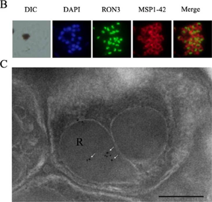
The expression and localization of PfRON3 in Plasmodium falciparum merozoites. (B) PfRON3 is expressed inside Plasmodium merozoites. Schizont stage parasites were dual-labeled with PfRON3-specific antibodies in combination with MSP1-42 antibodies. Nuclei are stained with DAPI. MSP1-42 protein was located at the merozoite membrane (red fluorescence) and RON3 was located inside merozoites (green fluorescence). (C) PfRON3 location shown via immunoelectronic microscopy. The section of merozoite in mature schizonts was labeled with purified rabbit anti-PfRON3 antibodies and subsequently detected with a secondary antibody conjugated with gold particles (arrow-headed). The black dots indicate signals from gold particles localized in the rhoptry body. Bar represents 200 nm.Zhao X, Chang Z, Tu Z, Yu S, Wei X, Zhou J, Lu H, Jiang N, Chen Q. PfRON3 is an erythrocyte-binding protein and a potential blood-stage vaccine candidate antigen. Malar J. 2014 Dec 12;13(1):490.
See original on MMP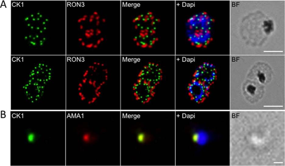
Localisation of PfCK1 in micronemes. (A) Localisation of PfCK1 and PfRON3 in schizonts and segmenters. DAPI was used to stain the nucleus and the scale bar represents 10μm. (B) Localisation of PfCK1 and AMA-1 in merozoites. DAPI was used to stain the nucleus and the scale bar represents 1μm. A punctate pattern emerges in segmenters, with one PfCK1 dot in each developing merozoite, adjacent to the rhoptry marker RON3. In line with this observation, PfCK1 colocalises with AMA-1, a protein of micronemes that is released at the very early stages of invasion.Dorin-Semblat D, Demarta-Gatsi C, Hamelin R, Armand F, Carvalho TG, Moniatte M, Doerig C. Malaria Parasite-Infected Erythrocytes Secrete PfCK1, the Plasmodium Homologue of the Pleiotropic Protein Kinase Casein Kinase 1. PLoS One. 2015 10(12):e0139591.
See original on MMPMore information
| PlasmoDB | PF3D7_1252100 |
| GeneDB | PF3D7_1252100 |
| Malaria Metabolic Pathways | Localisation images Pathways mapped to |
| Previous ID(s) | 2277.t00499, MAL12P1.496, PFL2505c |
| Orthologs | PBANKA_1464900 , PCHAS_1467100 , PKNH_1472100 , PVP01_1469200 , PVX_101485 , PY17X_1467600 |
| Google Scholar | Search for all mentions of this gene |