PF3D7_1240600 erythrocyte membrane protein 1, PfEMP1 (VAR)
Disruptability [+]
| Species | Disruptability | Reference | Submitter |
|---|---|---|---|
| P. falciparum 3D7 |
Possible |
USF piggyBac screen (Insert. mut.) | USF PiggyBac Screen |
Mutant phenotypes [+]
None reported yet. Please press the '+' button above to add one.Imaging data (from Malaria Metabolic Pathways)
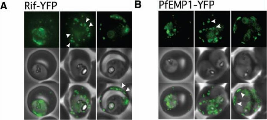
Export of Rifin surface antigens and of the major P. falciparum virulence factor, PfEMP1, depends on a functional Pexel motif. (A) A Rifin-YFP fusion including the Pexel motif is exported into the erythrocyte. Arrowheads indicate the localization of the reporter to punctate structures reminiscent of Maurer’s clefts (left panel, center) and later to the surface. (B) Export of PfEMP1-YFP also depends on a functional Pexel motif. As for the Rifin-YFP fusion, the reporter transiently localizes to structures similar to Maurer’s clefts, followed by a rim fluorescence suggestive of surface localization. Representative cells expressing the exported reporter are shown in ring stage (left), early (center), and late trophozoites (right). Marti M, Good RT, Rug M, Knuepfer E, Cowman AF. Targeting malaria virulence and remodeling proteins to the host erythrocyte. Science. 2004 306(5703):1930-3. Copyright 2010.
See original on MMP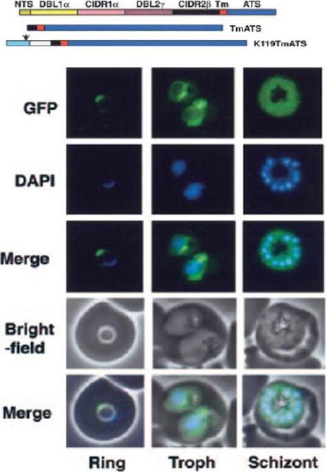
Expression of PfEMP1-GFP chimeras in P falciparum–infected RBCs. Fluorescence images of 3D7-TmATS at different stages.Spycher C, Rug M, Klonis N, Ferguson DJ, Cowman AF, Beck HP, Tilley L. Genesis of and trafficking to the Maurer's clefts of Plasmodium falciparum-infected erythrocytes. Mol Cell Biol. 2006 26:4074-85.
See original on MMP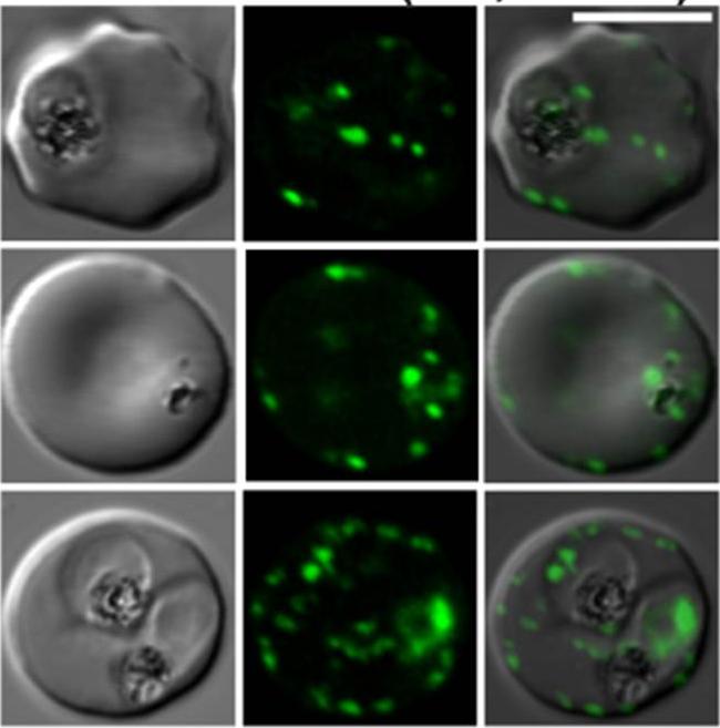
Confocal fluorescence microscopy images of transfected 3D7 P. falciparum-infected RBCs expressing a GFP chimeras directed to the Maurer’s clefts and red blood cell membrane. DIC image, the GFP fluorescence signal and an overlay of a transfectant expressing a fragment of P. falciparum erythrocyte membrane protein-1 in a chimeric construct: K119-EMP1-GFP. Scale bar = 5 µm. In trophozoite stage parasites K119-PfEMP1-GFP is associated with the Maurer’s clefts (top and middle rows). In more mature stages a weak rim signal is observed consistent with transfer of some of the K119-PfEMP1-GFP to the RBC membrane.Tilley L, McFadden G, Cowman A, Klonis N. Illuminating Plasmodium falciparum-infected red blood cells. Trends Parasitol. 2007 23:268-77.
See original on MMPMore information
| PlasmoDB | PF3D7_1240600 |
| GeneDB | PF3D7_1240600 |
| Malaria Metabolic Pathways | Localisation images Pathways mapped to |
| Previous ID(s) | 2277.t00392, MAL12P1.390, PFL1960w |
| Orthologs | |
| Google Scholar | Search for all mentions of this gene |