PF3D7_1235600 serine hydroxymethyltransferase (SHMT)
Disruptability [+]
| Species | Disruptability | Reference | Submitter | |
|---|---|---|---|---|
| P. falciparum 3D7 |
Refractory |
USF piggyBac screen (Insert. mut.) | USF PiggyBac Screen | |
| P. berghei ANKA |
Refractory |
RMgm-5223 | Imported from RMgmDB | |
Mutant phenotypes [+]
None reported yet. Please press the '+' button above to add one.Imaging data (from Malaria Metabolic Pathways)
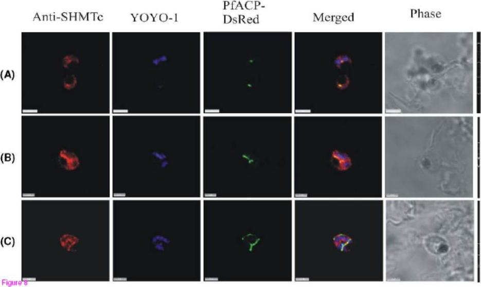
Positive control images using endogenously expressed DsRED-tagged ACP instead of anti-ACP antibodies. The use of only one primary antibody, anti-PfSHMTc (C-terminal end), with expressed DsRED tagged Pf ACP, was aimed at eliminating any possibility of artifactual fluorescence arising from interactions between two primary antibodies used simultaneously. (A) Two parasites, upper parasite is undergoing its first division, lower parasite is a late trophozoite. (B) Mitotic schizont with elongating apicoplast. (C) Mitotic schizont with ramifying apicoplast. All parasites show colocalization of anti-PfSHMTc fluorescence with the apicoplast, closely following the shape of the organelle, identical results to those obtained using two primary antibodies (scale bars (A) and (C) 3 μm, (B) 2 μm).Read M, Muller IB, Mitchell SL, Sims PF, Hyde JE. Dynamic subcellular localization of isoforms of the folate pathway enzyme serine hydroxymethyltransferase (SHMT) through the erythrocytic cycle of Plasmodium falciparum. Malar J. 2010 9:351.
See original on MMP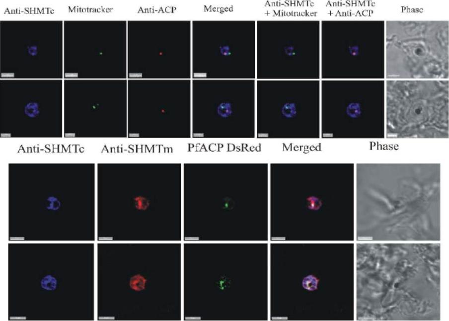
(A) and (B) Combined mitochondrial and apicoplast images probed with anti-PfSHMTc. These do not show nuclear morphology, therefore the erythrocytic cycle stage cannot be precisely ascertained; however, the size of the organelles and overall size of the parasites in (A) and (B) suggest that both are mid trophozoites. In (A) the parasite is probed with anti-PfSHMTc, MitoTracker and anti-ACP (plastid). The plastid is coincident with an area of marked PfSHMTc fluorescence, whereas the mitochondrion shows no evidence of coincident PfSHMTc fluorescence. In (B) the parasite is probed with anti-PfSHMTc, MitoTracker and anti-ACP (plastid). The plastid is coincident with a discrete area of PfSHMTc fluorescence, whereas the mitochondrion is located in a pocket of lower PfSHMTc fluorescence. (C) Parasite is probably a late trophozoite and (D) a mitotic schizont.. Both parasites were expressing DsRED-labelled ACP and were probed with both anti-PcSHMTc (IgY) and anti-PfSHMTm (IgG). The distribution of the two SHMT fluorescence signals are similar but not identical, and both co-localize with the apicoplast (scale bars (A) and (C), 3μm, (B) 2 μm, (D) 4 μm). Read M, Muller IB, Mitchell SL, Sims PF, Hyde JE. Dynamic subcellular localization of isoforms of the folate pathway enzyme serine hydroxymethyltransferase (SHMT) through the erythrocytic cycle of Plasmodium falciparum. Malar J. 2010 9:351
See original on MMP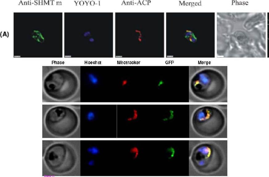
Upper panel: PfSHMTm (N-terminal) apicoplast immunofluorescent images illustrating the concentration of fluorescence in the extremities of elongating apicoplasts. (A) Mitotic schizont with an elongating apicoplast. Lower panel: GFP-tagging of truncated PfSHMTm in transfected 3D7 parasites. Fluorescence images of parasites transfected to yield a GFP-fusion carrying the first 100 amino acids of PfSHMTm at the N-terminus. MitoTracker was also used to localize the mitochondrion, which showed complete coincidence with the GFP fluorescence. YOYO- stains nuclei.Read M, Muller IB, Mitchell SL, Sims PF, Hyde JE. Dynamic subcellular localization of isoforms of the folate pathway enzyme serine hydroxymethyltransferase (SHMT) through the erythrocytic cycle of Plasmodium falciparum. Malar J. 2010 9:351.
See original on MMP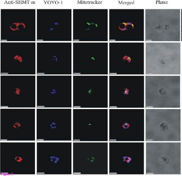
PfSHMTc immunofluorescence images showing localization in the mitochondrion. (A) Mid-trophozoite showing the association of a small mitochondrion with PfSHMTc fluorescence. (B) Early schizont showing association of an enlarged globular mitochondrion with a region of more intense PfSHMTc fluorescence. (C) Late schizont showing very little colocalization of mitochondria with areas of PfSHMTc fluorescence. Mitochondria are closely aligned to nuclei and show some co-localization with YOYO1 staining (scale bars 3μm).Read M, Muller IB, Mitchell SL, Sims PF, Hyde JE. Dynamic subcellular localization of isoforms of the folate pathway enzyme serine hydroxymethyltransferase (SHMT) through the erythrocytic cycle of Plasmodium falciparum. Malar J. 2010 9:351.
See original on MMP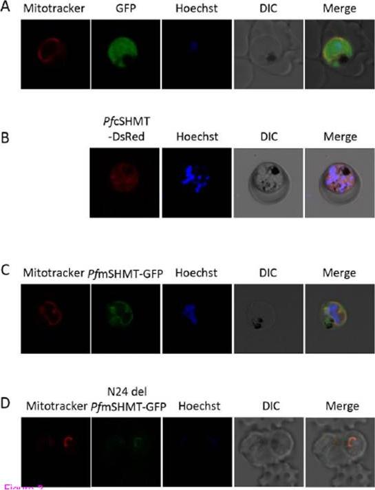
Localization of P. falciparum serine hydroxymethyltransferase (SHMT) isoforms. Parasites were transfected with pGFP (A), pRL_PfcSHMT PFL1720w (B), pGL_PfmSHMT PF14_0534 (C), and pGL_N24del PfmSHMT (D) plasmids expressing fluorescent signals from GFP or DsRed. Schematic diagrams of the recombinant plasmids used are shown alongside the confocal micrographs. Mitochondrion and nucleus is stained with Mitotracker™ (red) and Hoechst 33258 (blue) dye respectively. DIC; differential interference contrast. The distribution of DsRedtagged PfcSHMT appears to be dominantly in cytoplasm (B). GFP-tagged PfmSHMT was co-localized with Mitotracker™ within the mitochondria (C). N-24 truncated-PfmSHMTGFP with Mitotracker™ (D), indicating that this 24 N-terminal amino acid sequence does not play a role as a mitochondria targeting signal.Pornthanakasem W, Kongkasuriyachai D, Uthaipibull C, Yuthavong Y, Leartsakulpanich U. Plasmodium serine hydroxymethyltransferase: indispensability and display of distinct localization. Malar J. 2012 22;11(1):387.
See original on MMP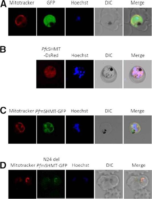
Localization of P. falciparum SHMT isoforms. Parasites were transfected with pGFP (A), pRL PfcSHMT (B), pGL_PfmSHMT (C), and pGL_N24del PfmSHMT (D) plasmids expressing fluorescent signals from GFP or DsRed. Schematic diagrams of the recombinant plasmids used are shown alongside the confocal micrographs. Mitochondrion and nucleus is stained with Mitotracker™ (red) and Hoechst 33258 (blue) dye respectively. DIC, differential interference contrast image. The distribution of DsRed-tagged PfcSHMT appears to be dominantly in cytoplasm (B). The distribution of GFP-tagged PfmSHMT was colocalized with Mitotracker™ within the mitochondria (C). Co-localization of N-24 truncated-PfmSHMTGFP with Mitotracker™ (D), indicated that this 24 N-terminal amino acid sequence does not play a role as a mitochondria targeting signal.Pornthanakasem W, Kongkasuriyachai D, Uthaipibull C, Yuthavong Y, Leartsakulpanich U. Plasmodium serine hydroxymethyltransferase: indispensability and display of distinct localization. Malar J. 2012 11:387.
See original on MMPMore information
| PlasmoDB | PF3D7_1235600 |
| GeneDB | PF3D7_1235600 |
| Malaria Metabolic Pathways | Localisation images Pathways mapped to |
| Previous ID(s) | 2277.t00344, MAL12P1.342, PFL1720w |
| Orthologs | PBANKA_1450200 , PCHAS_1452500 , PKNH_1455300 , PVP01_1453700 , PVX_100730 , PY17X_1452700 |
| Google Scholar | Search for all mentions of this gene |