PF3D7_1230700 protein transport protein SEC13 (SEC13)
Disruptability [+]
| Species | Disruptability | Reference | Submitter | |
|---|---|---|---|---|
| P. falciparum 3D7 |
Possible |
USF piggyBac screen (Insert. mut.) | USF PiggyBac Screen | |
| P. berghei ANKA |
Refractory |
PlasmoGEM (Barseq) | PlasmoGEM | |
Mutant phenotypes [+]
None reported yet. Please press the '+' button above to add one.Imaging data (from Malaria Metabolic Pathways)
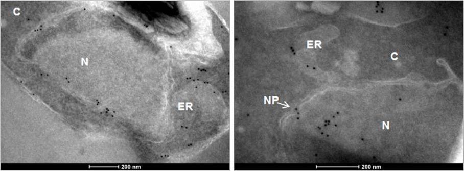
Immuno - EM of PfSec13-HA expressing trophozoites. PfSec13 localizes mainly to the nuclear envelope but gold particles can be also seen in the ER and cytosolic vesicles (left panel). In some nuclei PfSec13 is seen in the nucleoplasm (N) as well as in the nuclear pore (NP; white arrow) and the ER (right panel). (C; cytoplasm).Dahan-Pasternak N, Nasereddin A, Kolevzon N, Pe'er M, Wong W, Shinder V, Turnbull L, Whitchurch CB, Elbaum M, Gilberger TW, Yavin E, Baum J, Dzikowski R. PfSec13 is an unusual chromatin associated nucleoporin of Plasmodium falciparum, which is essential for parasite proliferation in human erythrocytes. J Cell Sci. 2013 126(Pt 14):3055-69 Reproduced with permission.
See original on MMP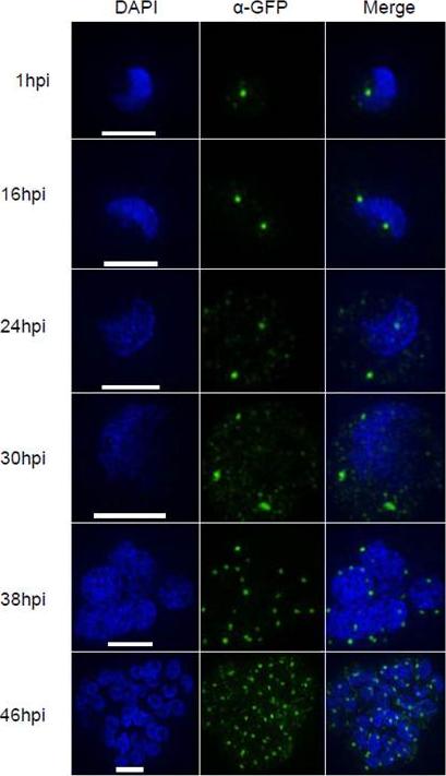
Dynamics of PfSec13-GFP during P. falciparum intra erythrocytic development. High resolution microscopy (3D-SIM) of PfSec13-GFP showing an increase in the number of foci as parasites develop into trophozoites (1- 30 hpi) and a gradual decrease during schizogony (36 and 48 hpi). The high resolution of these images enables differentiation between the large strong signals and the fainter ones. The size bar represents 2 μm for 1, 16, 24, 38 & 48 hpi and 1 μm for 30 hpi. Using super resolution microscopy it is possible to differentiate between strong signals at the nuclear periphery and weaker signals seen in the cytoplasm.Dahan-Pasternak N, Nasereddin A, Kolevzon N, Pe'er M, Wong W, Shinder V, Turnbull L, Whitchurch CB, Elbaum M, Gilberger TW, Yavin E, Baum J, Dzikowski R. PfSec13 is an unusual chromatin associated nucleoporin of Plasmodium falciparum, which is essential for parasite proliferation in human erythrocytes. J Cell Sci. 2013 126(Pt 14):3055-69 Reproduced with permission.
See original on MMP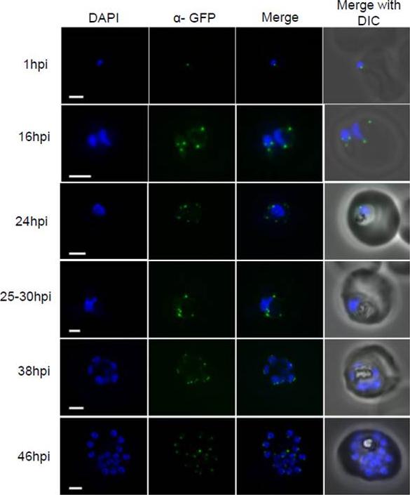
Dynamics of PfSec13-GFP during P. falciparum intra erythrocytic development. Conventional fluorescent imaging of PfSec13-GFP (Green) at different time points (hpi = hours post invasion) during the IDC. Strong signals of PfSec13-GFP at the periphery of the DAPI stained nucleus corresponds with NPC clusters. Dynamics in cluster number and orientation were documented from 1 hpi to 46 hpi. At the late schizont stage (from 38 hpi to 46 hpi) there is a change in NPC orientation. The size bar represents 1μm. The size bar represents 2 μm for 1, 16, 24, 38 & 48 hpi and 1 μm for 30 hpi. Strong signals are seen at the nuclear periphery and faint background in the cytoplasm. Dahan-Pasternak N, Nasereddin A, Kolevzon N, Pe'er M, Wong W, Shinder V, Turnbull L, Whitchurch CB, Elbaum M, Gilberger TW, Yavin E, Baum J, Dzikowski R. PfSec13 is an unusual chromatin associated nucleoporin of Plasmodium falciparum, which is essential for parasite proliferation in human erythrocytes. J Cell Sci. 2013 126(Pt 14):3055-69 Reproduced with permission.
See original on MMP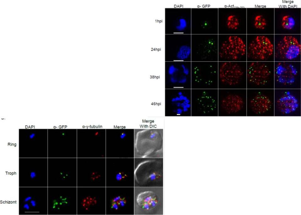
Right: PfSec13 is associated with γ-tubulin rather than with F-actin. Super resolution IFAs of the PfSec13-GFP (green) expressing parasites that were co-stained with F-actin (red) using rabbit α- Act239-253. Nuclei were stained with DAPI (blue). The size bar represents 1μm. (B), IFAs showing co-localization of PfSec13–GFP (green) and γ-tubulin (red) during the IDC. later in the IDC PfSec13 remains located at the nuclear periphery while F-actin is spread throughout the entire cell .Left: PfSec13 is associated with γ-tubulin rather than with F-actin. IFAs showing co-localization of PfSec13–GFP (green) and γ-tubulin (red) during the IDC. PfSec13 is associated with F-actin at the nuclear periphery. The association of PfSec13 with γ-tubulin may indicate a possible role of microtubules in NPCs dynamics.Dahan-Pasternak N, Nasereddin A, Kolevzon N, Pe'er M, Wong W, Shinder V, Turnbull L, Whitchurch CB, Elbaum M, Gilberger TW, Yavin E, Baum J, Dzikowski R. PfSec13 is an unusual chromatin associated nucleoporin of Plasmodium falciparum, which is essential for parasite proliferation in human erythrocytes. J Cell Sci. 2013 126(Pt 14):3055-69
See original on MMP
Right: PfSec13 does not co-localize with P. falciparum heterochromatin. IFAs of the PfSec13-GFP expressing parasites that were co-immunolabled with α-GFP (green) and α-H3K9me3 (red) at different stages of the IDC. In 85% of the counted nuclei of all stages (n=100), the H3K9me3 signal and PfSec13-GFP did not co-localize.Left: In vivo imaging of parasites expressing both PfSec13-GFP and PfHP1-HA-mCherry during the IDC. Although PfSec13 (green) and PfHP1 (red) localized to the nuclear periphery they do not co-localize.Righet lower corner: In vivo confocal microscopy of parasites expressing both PfSec13-GFP and PfHP1-HA-mCherry.Dahan-Pasternak N, Nasereddin A, Kolevzon N, Pe'er M, Wong W, Shinder V, Turnbull L, Whitchurch CB, Elbaum M, Gilberger TW, Yavin E, Baum J, Dzikowski R. PfSec13 is an unusual chromatin associated nucleoporin of Plasmodium falciparum, which is essential for parasite proliferation in human erythrocytes. J Cell Sci. 2013 126(Pt 14):3055-69 Reproduced with permission
See original on MMP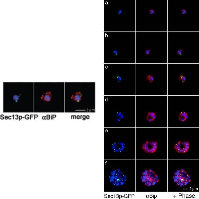
Right: Comparative analyses of transitional ER (tER) dynamics (PfSec13p) with the ER throughout the asexual life cycle of P. falciparum. Transgenic parasites expressing Sec13p–GFP (green) were fixed and stained with antibodies against the ER marker BiP (red). In ring-stage parasites (8-16 hours post invasion) GFP fluorescence is restricted to a tightly defined area in close proximity to the nucleus (a-b). A multiplication of the Sec13p–GFP-defined ER exit site takes place prior to nuclear division (24 hours post invasion, c). As the parasite matures nuclear division commences and the ER forms a more complex network, which is accompanied by further tER site multiplication (d-f).Left: Colocalization of Sec13p–GFP (green) with the ER marker BiP (red) in fixed parasites. Antibodies against the luminal chaperone visualize the ER as a nuclear envelope with some protrusions. Sec13p–GFP is restricted to defined regions within the ER (merged image). All images show the nucleus in blue (DAPI).Struck NS, Herrmann S, Schmuck-Barkmann I, de Souza Dias S, Haase S, Cabrera AL, Treeck M, Bruns C, Langer C, Cowman AF, Marti M, Spielmann T, Gilberger TW. Spatial dissection of the cis- and trans-Golgi compartments in the malaria parasite Plasmodium falciparum. Mol Microbiol. 2008 67:1320-30.
See original on MMP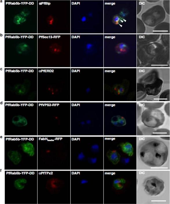
Localization of PfRab5b to a punctate compartment in the parasite cytoplasm. Triple staining with PfRab5b-YFP-DD (green), DAPI (blue) and one of the following markers (red): PfBip (a, ER), PfSec13-RFP (b, ER exit site), PfERD2 (c, Golgi), PfVPS2-RFP (d, putative multivesicular body/endosome), FabHleader-RFP (e, apicoplast), or PfTPx-2 (f, mitochondria) after 24 h incubation with Shld1. PfRab5b-YFP-DD localized adjacent to the Bip signal (arrowheads). Bars 5 μm.Ebine K, Hirai M, Sakaguchi M, Yahata K, Kaneko O, Saito-Nakano Y. Plasmodium Rab5b is secreted to the cytoplasmic face of the tubovesicular network in infected red blood cells together with N-acylated adenylate kinase 2. Malar J. 2016 15:323.
See original on MMPMore information
| PlasmoDB | PF3D7_1230700 |
| GeneDB | PF3D7_1230700 |
| Malaria Metabolic Pathways | Localisation images Pathways mapped to |
| Previous ID(s) | 2277.t00296, MAL12P1.296, PFL1480w |
| Orthologs | PBANKA_1445400 , PCHAS_1447600 , PKNH_1450100 , PVP01_1448900 , PVX_124175 , PY17X_1447900 |
| Google Scholar | Search for all mentions of this gene |