PF3D7_1230400 ATP-dependent protease subunit ClpQ (ClpQ)
Disruptability [+]
No information reported yet. Please press the '+' button above to add some.Mutant phenotypes [+]
| Species | Stage | Phenotype | Reference | Submitter |
|---|---|---|---|---|
| P. falciparum 3D7 | Asexual |
Difference from wild-type |
23521916 (Conditional)
\" A proteolytically inactive mutant PfClpQ protein [PfClpQ(mut)] fused with FKBP degradation domain was expressed in parasites, which gets stabilized by Shield1 drug treatment. We show that the inactive PfClpQ(mut) interacts with wild-type PfClpQ and associates within multi-subunit complex in the parasite. Stabilization of the PfClpQ(mut) and its association in the protease machinery caused dominant negative effect in the transgenic parasites, which disrupted the growth cycle of asexual blood stage parasites. The mitochondria in these parasites showed abnormal morphology, these mitochondria were not able to grow and divide in the parasite. We further show that the dominant negative effect of PfClpQ(mut) disrupted transcription of mitochondrial genome encoded genes, which in turn blocked normal development and functioning of the mitochondria.\" |
Theo Sanderson, Francis Crick Institute |
Imaging data (from Malaria Metabolic Pathways)
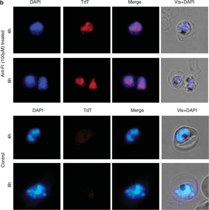
DNA fragmentation in P. falciparum parasites as assessed by TUNEL staining. Fluorescent microscopic images of TUNEL-positive parasites (TdT red staining) in treated cultures as compared with controls. The parasite nuclei were stained with DAPI (blue) and parasites were visualized by fluorescence microscope. Dl is a synthetic peptide that binds PfClpQ, thereby revealing it location. ClpQ protease is located in the matrix of the mitochondrion.Rathore S, Jain S, Sinha D, Gupta M, Asad M, Srivastava A, Narayanan MS, Ramasamy G, Chauhan VS, Gupta D, Mohmmed A. Disruption of a mitochondrial protease machinery in Plasmodium falciparum is an intrinsic signal for parasite cell death. Cell Death Dis. 2011 24;2:e231.
See original on MMP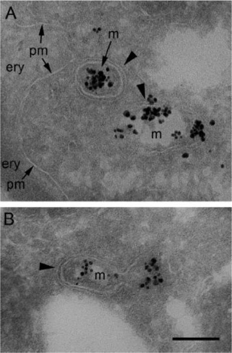
Ultrathin immunogold-labeled cryosections of Plasmodium falciparum HslV (PfHslV)-enhanced yellow fluorescent protein (EYFP) expressing parasites residing in erythrocytes (ery). Mitochondria (m) are labeled with anti-GFP antibodies and silver-enhanced Nanogold. (A) Two mitochondria close to the parasite’s plasma membrane (pm). (B) A longitudinal section through a gold decorated mitochondrion. Arrowheads point to double membranes of mitochondria. Gold labeling on non-transfected parasites was negligible. Bar = 200 nm.Tschan S, Kreidenweiss A, Stierhof YD, Sessler N, Fendel R, Mordmüller B. Mitochondrial localization of the threonine peptidase PfHslV, a ClpQ ortholog in Plasmodium falciparum. Int J Parasitol. 2010 40:1517-23 Copyright Elsevier 2010
See original on MMP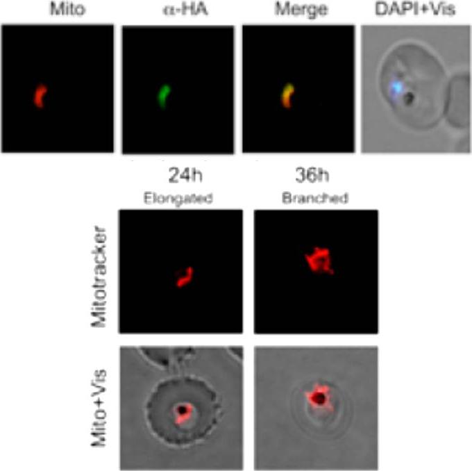
Upper panel: Immunofluorescence assay showing transgenic parasites expressing PfClpQ(mut)-HA-ddFKBP (ClpQ(mut)-DD parasite line) immuno-stained with anti-HA antibodies and co-stained for mitochondria with MitoTracker. The parasite nuclei were stained with DAPI and slides were visualized by fluorescence microscope. the immunofluorescence staining overlapped with the MitoTracker staining suggesting that the fusion protein is localized in the mitochondria which is the site of action of native proteinLower panel: Fluorescent microscopic images of MitoTracker stained ClpQ(mut)-DD transgenic parasites at different time points (lower panel). The mitochondria in control ClpQ(mut)-DD set showed normal growth and development; in majority of these parasites at trophozoite stage the mitochondria appeared as elongated structures, in late trophozoite/early schizont stage) most of these mitochondria showed branched structure.Jain S, Rathore S, Asad M, Hossain ME, Sinha D, Datta G, Mohmmed A. The prokaryotic ClpQ protease plays a key role in growth and development of mitochondria in Plasmodium falciparum. Cell Microbiol. 2013 15(10):1660-73
See original on MMP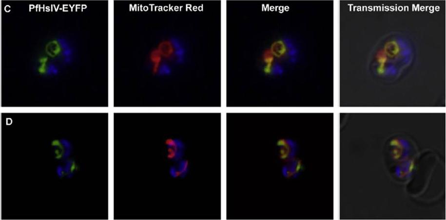
Immunofluorescence and fluorescence microscopy on Plasmodium falciparum HslV (PfHslV)-enhanced yellow fluorescent protein (EYFP) expressing parasites. C and D: fluorescence microscopy using MitoTracker Red CMXRos (red) and Hoechst 33342 (blue). Bar = 2 mm. D10_PfHslV-EYFP late-stage parasites showed a distinct fluorescence pattern reminiscent of an organellar structure. PfHslV-EYFP is located in the plasmodial mitochondrion.Tschan S, Kreidenweiss A, Stierhof YD, Sessler N, Fendel R, Mordmüller B. Mitochondrial localization of the threonine peptidase PfHslV, a ClpQ ortholog in Plasmodium falciparum. Int J Parasitol. 2010 40:1517-23 Copyright Elsevier 2010
See original on MMP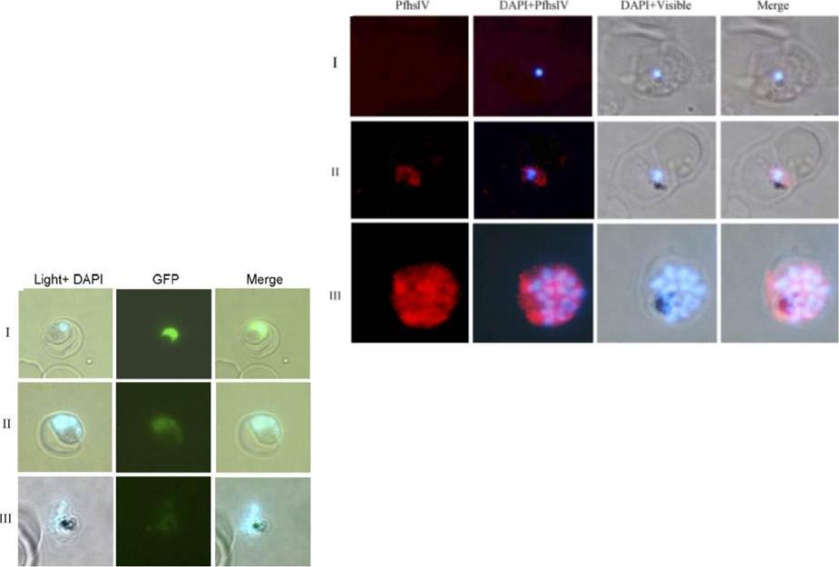
Left: Immuno-fluorescence assay to localize PfHslV in the parasite: thin parasite smears at ring (panel I), trophozoite (panel II) or schizont (panel III) stages were fixed and immunostained with anti-PfHslV (green) antibodies followed by Cy3 labeled secondary antibodies. The parasite nuclei were stained with DAPI (blue) and slides were visualized by fluorescence microscope. In both trophozoite and schizont stages, fluorescence was distributed specifically in the parasite cytoplasm. Left: Activity of native PfHslV in the parasite: P. falciparum parasites were transfected with transfection vector construct containing Arc-GFP chimeric gene under control of constitutive promoter hsp86. (A) Fluorescent microscopic images of transgenic parasites at ring (panel I), trophozoite (panel II) and schizont (panel III) stages showing differential level of GFP fluorescence. Parasite nuclei are stained with DAPI (blue).Ramasamy G, Gupta D, Mohmmed A, Chauhan VS. Characterization and localization of Plasmodium falciparum homolog of prokaryotic ClpQ/HslV protease. Mol Biochem Parasitol. 2007 152(2):139-48.
See original on MMPMore information
| PlasmoDB | PF3D7_1230400 |
| GeneDB | PF3D7_1230400 |
| Malaria Metabolic Pathways | Localisation images Pathways mapped to |
| Previous ID(s) | 2277.t00293, MAL12P1.293, PFL1465c |
| Orthologs | PBANKA_1445100 , PCHAS_1447300 , PKNH_1449800 , PVP01_1448600 , PVX_124160 , PY17X_1447600 |
| Google Scholar | Search for all mentions of this gene |